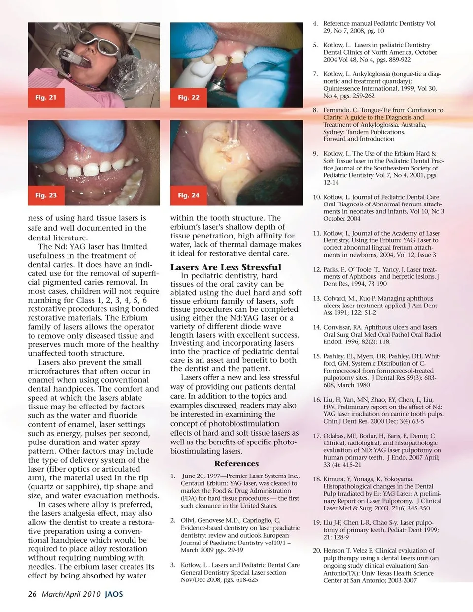
4. Reference manual Pediatric Dentistry Vol 29, No 7, 2008, pg. 10 5. Kotlow, L. Lasers in pediatric Dentistry Dental Clinics of North America, October 2004 Vol 48, No 4, pgs. 889-922 Fig. 21 Fig. 22 7. Kotlow, L. Ankyloglossia (tongue-tie a diag- nostic and treatment quandary); Quintessence International, 1999, Vol 30, No 4, pgs. 259-262 8. Fernando, C. Tongue-Tie from Confusion to Clarity. A guide to the Diagnosis and Treatment of Ankyloglossia. Australia, Sydney: Tandem Publications. Forward and Introduction 9. Kotlow, L. The Use of the Erbium Hard & Soft Tissue laser in the Pediatric Dental Prac- tice Journal of the Southeastern Society of Pediatric Dentistry Vol 7, No 4, 2001, pgs. 12-14 Fig. 23 ness of using hard tissue lasers is safe and well documented in the dental literature. The Nd: YAG laser has limited usefulness in the treatment of dental caries. It does have an indi- cated use for the removal of superfi- cial pigmented caries removal. In most cases, children will not require numbing for Class 1, 2, 3, 4, 5, 6 restorative procedures using bonded restorative materials. The Erbium family of lasers allows the operator to remove only diseased tissue and preserves much more of the healthy unaffected tooth structure. Lasers also prevent the small microfractures that often occur in enamel when using conventional dental handpieces. The comfort and speed at which the lasers ablate tissue may be effected by factors such as the water and fluoride content of enamel, laser settings such as energy, pulses per second, pulse duration and water spray pattern. Other factors may include the type of delivery system of the laser (fiber optics or articulated arm), the material used in the tip (quartz or sapphire), tip shape and size, and water evacuation methods. In cases where alloy is preferred, the lasers analgesia effect, may also allow the dentist to create a restora- tive preparation using a conven- tional handpiece which would be required to place alloy restoration without requiring numbing with needles. The erbium laser creates its effect by being absorbed by water 26 March/April 2010 JAOS Fig. 24 within the tooth structure. The erbium’s laser’s shallow depth of tissue penetration, high affinity for water, lack of thermal damage makes it ideal for restorative dental care. Lasers Are Less Stressful In pediatric dentistry, hard tissues of the oral cavity can be ablated using the duel hard and soft tissue erbium family of lasers, soft tissue procedures can be completed using either the Nd:YAG laser or a variety of different diode wave length lasers with excellent success. Investing and incorporating lasers into the practice of pediatric dental care is an asset and benefit to both the dentist and the patient. Lasers offer a new and less stressful way of providing our patients dental care. In addition to the topics and examples discussed, readers may also be interested in examining the concept of photobiostimulation effects of hard and soft tissue lasers as well as the benefits of specific photo- biostimulating lasers. References 1. June 20, 1997—Premier Laser Systems Inc., Centauri Erbium: YAG laser, was cleared to market the Food & Drug Administration (FDA) for hard tissue procedures — the first such clearance in the United States. 2. Olivi, Genovese M.D., Caprioglio, C. Evidence-based dentistry on laser peadiatric dentistry: review and outlook European Journal of Paediatric Dentistry vol10/1 – March 2009 pgs. 29-39 3. Kotlow, L . Lasers and Pediatric Dental Care General Dentistry Special Laser section Nov/Dec 2008, pgs. 618-625 10. Kotlow, L. Journal of Pediatric Dental Care Oral Diagnosis of Abnormal frenum attach- ments in neonates and infants, Vol 10, No 3 October 2004 11. Kotlow, L. Journal of the Academy of Laser Dentistry, Using the Erbium: YAG Laser to correct abnormal lingual frenum attach- ments in newborns, 2004, Vol 12, Issue 3 12. Parks, F., O’ Toole, T., Yancy, J. Laser treat- ments of Aphthous and herpetic lesions. J Dent Res, 1994, 73 190 13. Colvard, M., Kuo P. Managing aphthous ulcers; laser treatment applied. J Am Dent Ass 1991; 122: 51-2 14. Convissar, RA. Aphthous ulcers and lasers. Oral Surg Oral Med Oral Pathol Oral Radiol Endod. 1996; 82(2): 118. 15. Pashley, EL, Myers, DR, Pashley, DH, Whit- ford, GM. Systemic Distribution of C- Formocreosol from formocreosol-treated pulpotomy sites. J Dental Res 59(3): 603- 608, March 1980 16. Liu, H, Yan, MN, Zhao, EY, Chen, L, Liu, HW. Preliminary report on the effect of Nd: YAG laser irradiation on canine tooth pulps. Chin J Dent Res. 2000 Dec; 3(4) 63-5 17. Odabas, ME, Bodur, H, Baris, E, Demir, C Clinical, radiological, and histopathologic evaluation of ND: YAG laser pulpotomy on human primary teeth. J Endo, 2007 April; 33 (4): 415-21 18. Kimura, Y, Yonaga, K, Yokoyama. Histopathological changes in the Dental Pulp Irradiated by Er: YAG Laser: A prelimi- nary Report on Laser Pulpotomy. J Clinical Laser Med & Surg. 2003, 21(6) 345-350 19. Liu J-F, Chen L-R, Chao S-y. Laser pulpo- tomy of primary teeth. Pediatr Dent 1999; 21: 128-9 20. Henson T. Velez E. Clinical evaluation of pulp therapy using a dental lasers unit (an ongoing study clinical evaluation) San Antonio(TX): Univ Texas Health Science Center at San Antonio; 2003-2007
Journal of the American Orthodontic Society March - April 2010: Page 26