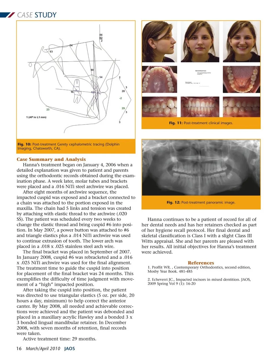
CASE STUDY Fig. 11: Post-treatment clinical images. Fig. 10: Post-treatment Gerety cephalometric tracing (Dolphin Imaging, Chatsworth, CA). Case Summary and Analysis Hanna’s treatment began on January 4, 2006 when a detailed explanation was given to patient and parents using the orthodontic records obtained during the exam- ination phase. A week later, molar tubes and brackets were placed and a .016 NiTi steel archwire was placed. After eight months of archwire sequence, the impacted cuspid was exposed and a bracket connected to a chain was attached to the portion exposed in the maxilla. The chain had 5 links and tension was created by attaching with elastic thread to the archwire (.020 SS). The patient was scheduled every two weeks to change the elastic thread and bring cuspid #6 into posi- tion. In May 2007, a power button was attached to #6 and triangle elastics plus a .014 NiTi archwire was used to continue extrusion of tooth. The lower arch was placed in a .018 x .025 stainless steel arch wire. The final bracket was placed in September of 2007. In January 2008, cuspid #6 was rebracketed and a .016 x .025 NiTi archwire was used for the final alignment. The treatment time to guide the cuspid into position for placement of the final bracket was 24 months. This exemplifies the difficulty of time judgment with move- ment of a “high” impacted position. After taking the cuspid into position, the patient was directed to use triangular elastics (5 oz. per side, 20 hours a day, minimum) to help correct the anterior canter. By May 2008, all needed and achievable correc- tions were achieved and the patient was debonded and placed in a maxillary acrylic Hawley and a bonded 3 x 3 bonded lingual mandibular retainer. In December 2008, with seven months of retention, final records were taken. Active treatment time: 29 months. 16 March/April 2010 JAOS Fig. 12: Post-treatment panoramic image. Hanna continues to be a patient of record for all of her dental needs and has her retainers checked as part of her hygiene recall protocol. Her final dental and skeletal classification is Class I with a slight Class III Witts appraisal. She and her parents are pleased with her results. All initial objectives for Hanna’s treatment were achieved. References 1. Proffit WR. , Contemporary Orthodontics, second edition, Mosby Year Book. 481-485 2. Echeverri JC., Impacted incisors in mixed dentition. JAOS, 2009 Spring Vol 9 (1): 16-20
Journal of the American Orthodontic Society March - April 2010: Page 16