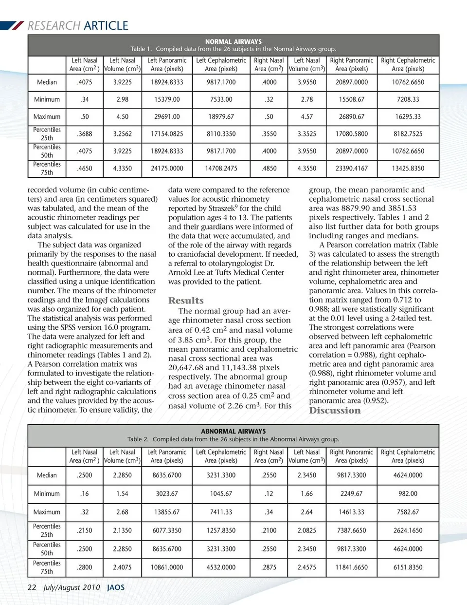
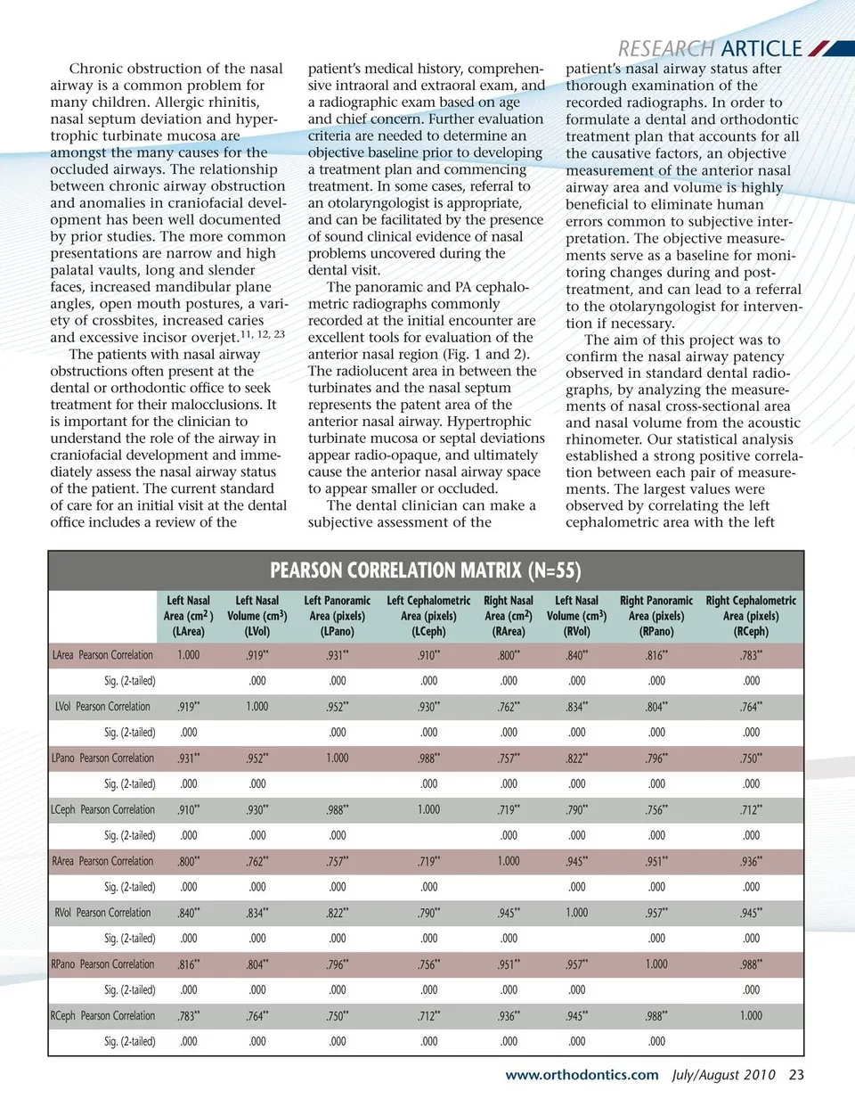
RESEARCH ARTICLE Chronic obstruction of the nasal airway is a common problem for many children. Allergic rhinitis, nasal septum deviation and hyper-trophic turbinate mucosa are amongst the many causes for the occluded airways. The relationship between chronic airway obstruction and anomalies in craniofacial devel-opment has been well documented by prior studies. The more common presentations are narrow and high palatal vaults, long and slender faces, increased mandibular plane angles, open mouth postures, a vari-ety of crossbites, increased caries and excessive incisor overjet.11, 12, 23 The patients with nasal airway obstructions often present at the dental or orthodontic office to seek treatment for their malocclusions. It is important for the clinician to understand the role of the airway in craniofacial development and imme-diately assess the nasal airway status of the patient. The current standard of care for an initial visit at the dental office includes a review of the patient’s medical history, comprehen-sive intraoral and extraoral exam, and a radiographic exam based on age and chief concern. Further evaluation criteria are needed to determine an objective baseline prior to developing a treatment plan and commencing treatment. In some cases, referral to an otolaryngologist is appropriate, and can be facilitated by the presence of sound clinical evidence of nasal problems uncovered during the dental visit. The panoramic and PA cephalo-metric radiographs commonly recorded at the initial encounter are excellent tools for evaluation of the anterior nasal region (Fig. 1 and 2). The radiolucent area in between the turbinates and the nasal septum represents the patent area of the anterior nasal airway. Hypertrophic turbinate mucosa or septal deviations appear radio-opaque, and ultimately cause the anterior nasal airway space to appear smaller or occluded. The dental clinician can make a subjective assessment of the patient’s nasal airway status after thorough examination of the recorded radiographs. In order to formulate a dental and orthodontic treatment plan that accounts for all the causative factors, an objective measurement of the anterior nasal airway area and volume is highly beneficial to eliminate human errors common to subjective inter-pretation. The objective measure-ments serve as a baseline for moni-toring changes during and post-treatment, and can lead to a referral to the otolaryngologist for interven-tion if necessary. The aim of this project was to confirm the nasal airway patency observed in standard dental radio-graphs, by analyzing the measure-ments of nasal cross-sectional area and nasal volume from the acoustic rhinometer. Our statistical analysis established a strong positive correla-tion between each pair of measure-ments. The largest values were observed by correlating the left cephalometric area with the left PEARSON CORRELATION MATRIX (N=55) Left Nasal Area (cm2 ) (LArea) LArea Pearson Correlation Sig. (2-tailed) LVol Pearson Correlation Sig. (2-tailed) LPano Pearson Correlation Sig. (2-tailed) LCeph Pearson Correlation Sig. (2-tailed) RArea Pearson Correlation Sig. (2-tailed) RVol Pearson Correlation Sig. (2-tailed) RPano Pearson Correlation Sig. (2-tailed) RCeph Pearson Correlation Sig. (2-tailed) 1.000 .919** .000 .931** .000 .910** .000 .800** .000 .840** .000 .816** .000 .783** .000 Left Nasal Volume (cm3) (LVol) .919** .000 1.000 .952** .000 .930** .000 .762** .000 .834** .000 .804** .000 .764** .000 Left Panoramic Area (pixels) (LPano) .931** .000 .952** .000 1.000 .988** .000 .757** .000 .822** .000 .796** .000 .750** .000 Left Cephalometric Area (pixels) (LCeph) .910** .000 .930** .000 .988** .000 1.000 .719** .000 .790** .000 .756** .000 .712** .000 Right Nasal Area (cm2) (RArea) .800** .000 .762** .000 .757** .000 .719** .000 1.000 .945** .000 .951** .000 .936** .000 Left Nasal Volume (cm3) (RVol) .840** .000 .834** .000 .822** .000 .790** .000 .945** .000 1.000 .957** .000 .945** .000 Right Panoramic Area (pixels) (RPano) .816** .000 .804** .000 .796** .000 .756** .000 .951** .000 .957** .000 1.000 .988** .000 Right Cephalometric Area (pixels) (RCeph) .783** .000 .764** .000 .750** .000 .712** .000 .936** .000 .945** .000 .988** .000 1.000 www.orthodontics.com July/August 2010 23
Journal of the American Orthodontic Society July-August 2010/Buyer's Guide: Page 23