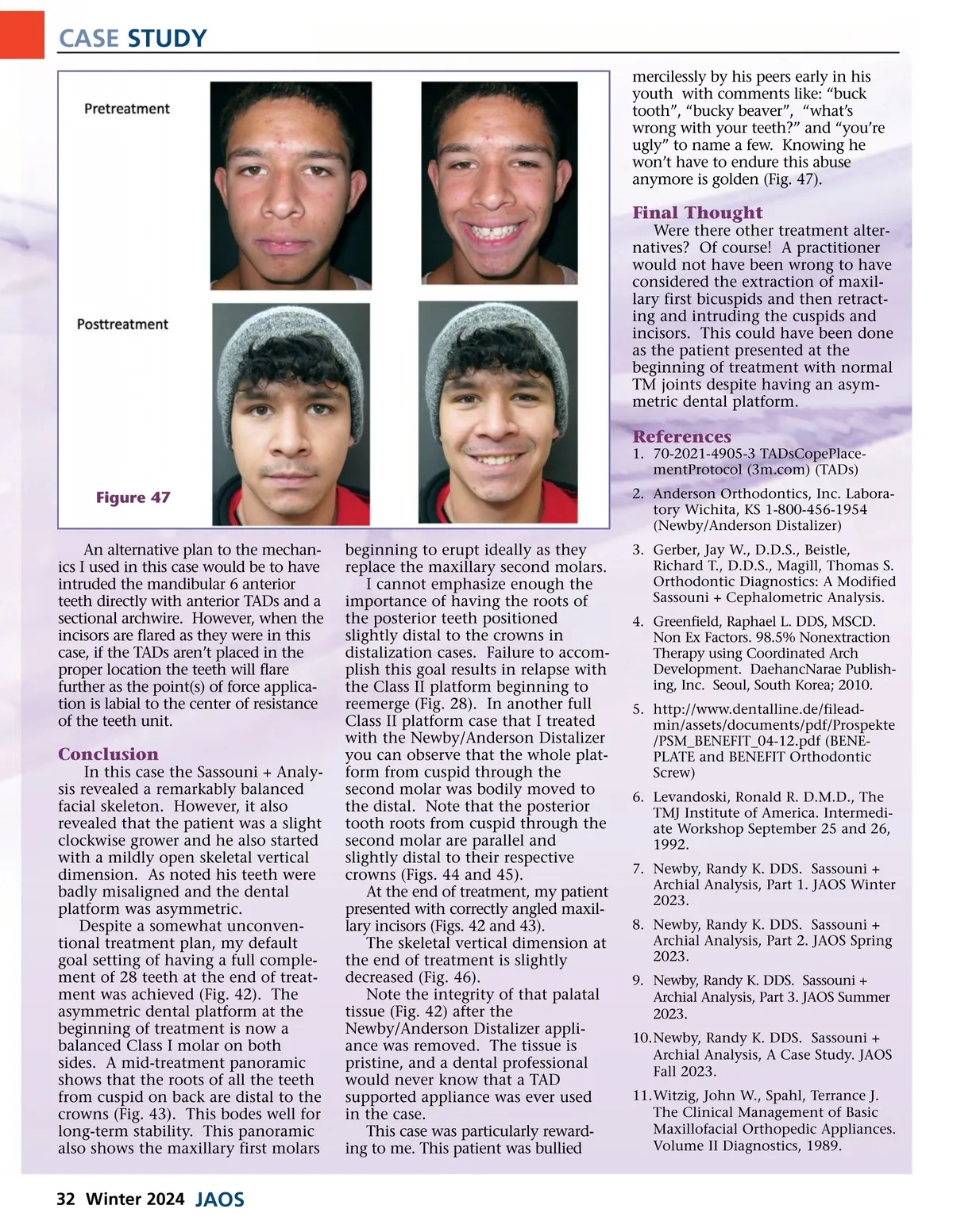
CASE STUDY mercilessly by his peers early in his youth with comments like: “buck tooth”, “bucky beaver”, “what’s wrong with your teeth?” and “you’re ugly” to name a few. Knowing he won’t have to endure this abuse anymore is golden (Fig. 47). Final Thought Were there other treatment alter-natives? Of course! A practitioner would not have been wrong to have considered the extraction of maxil-lary first bicuspids and then retract-ing and intruding the cuspids and incisors. This could have been done as the patient presented at the beginning of treatment with normal TM joints despite having an asym-metric dental platform. References 1. 70-2021-4905-3 TADsCopePlace-mentProtocol (3m.com) (TADs) 2. Anderson Orthodontics, Inc. Labora-tory Wichita, KS 1-800-456-1954 (Newby/Anderson Distalizer) Figure 47 An alternative plan to the mechan-ics I used in this case would be to have intruded the mandibular 6 anterior teeth directly with anterior TADs and a sectional archwire. However, when the incisors are flared as they were in this case, if the TADs aren’t placed in the proper location the teeth will flare further as the point(s) of force applica-tion is labial to the center of resistance of the teeth unit. beginning to erupt ideally as they replace the maxillary second molars. I cannot emphasize enough the importance of having the roots of the posterior teeth positioned slightly distal to the crowns in distalization cases. Failure to accom-plish this goal results in relapse with the Class II platform beginning to reemerge (Fig. 28). In another full Class II platform case that I treated with the Newby/Anderson Distalizer you can observe that the whole plat-form from cuspid through the second molar was bodily moved to the distal. Note that the posterior tooth roots from cuspid through the second molar are parallel and slightly distal to their respective crowns (Figs. 44 and 45). At the end of treatment, my patient presented with correctly angled maxil-lary incisors (Figs. 42 and 43). The skeletal vertical dimension at the end of treatment is slightly decreased (Fig. 46). Note the integrity of that palatal tissue (Fig. 42) after the Newby/Anderson Distalizer appli-ance was removed. The tissue is pristine, and a dental professional would never know that a TAD supported appliance was ever used in the case. This case was particularly reward-ing to me. This patient was bullied 3. Gerber, Jay W., D.D.S., Beistle, Richard T., D.D.S., Magill, Thomas S. Orthodontic Diagnostics: A Modified Sassouni + Cephalometric Analysis. 4. Greenfield, Raphael L. DDS, MSCD. Non Ex Factors. 98.5% Nonextraction Therapy using Coordinated Arch Development. DaehancNarae Publish-ing, Inc. Seoul, South Korea; 2010. 5. http://www.dentalline.de/filead-min/assets/documents/pdf/Prospekte /PSM_BENEFIT_04-12.pdf (BENE-PLATE and BENEFIT Orthodontic Screw) 6. Levandoski, Ronald R. D.M.D., The TMJ Institute of America. Intermedi-ate Workshop September 25 and 26, 1992. 7. Newby, Randy K. DDS. Sassouni + Archial Analysis, Part 1. JAOS Winter 2023. 8. Newby, Randy K. DDS. Sassouni + Archial Analysis, Part 2. JAOS Spring 2023. 9. Newby, Randy K. DDS. Sassouni + Archial Analysis, Part 3. JAOS Summer 2023. 10.Newby, Randy K. DDS. Sassouni + Archial Analysis, A Case Study. JAOS Fall 2023. 11.Witzig, John W., Spahl, Terrance J. The Clinical Management of Basic Maxillofacial Orthopedic Appliances. Volume II Diagnostics, 1989. Conclusion In this case the Sassouni + Analy-sis revealed a remarkably balanced facial skeleton. However, it also revealed that the patient was a slight clockwise grower and he also started with a mildly open skeletal vertical dimension. As noted his teeth were badly misaligned and the dental platform was asymmetric. Despite a somewhat unconven-tional treatment plan, my default goal setting of having a full comple-ment of 28 teeth at the end of treat-ment was achieved (Fig. 42). The asymmetric dental platform at the beginning of treatment is now a balanced Class I molar on both sides. A mid-treatment panoramic shows that the roots of all the teeth from cuspid on back are distal to the crowns (Fig. 43). This bodes well for long-term stability. This panoramic also shows the maxillary first molars 32 Winter 2024 JAOS
Journal of the American Orthodontic Society JAOS Winter 2024: Page 32