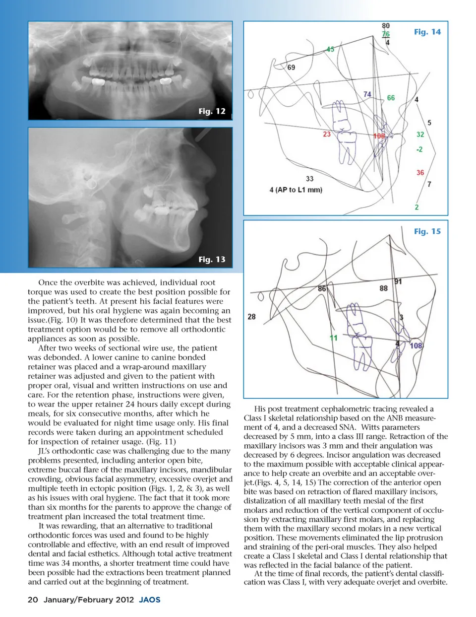
Fig. 14 Fig. 12 Fig. 15 Fig. 13 Once the overbite was achieved, individual root torque was used to create the best position possible for the patient’s teeth. At present his facial features were improved, but his oral hygiene was again becoming an issue.(Fig. 10) It was therefore determined that the best treatment option would be to remove all orthodontic appliances as soon as possible. After two weeks of sectional wire use, the patient was debonded. A lower canine to canine bonded retainer was placed and a wrap-around maxillary retainer was adjusted and given to the patient with proper oral, visual and written instructions on use and care. For the retention phase, instructions were given, to wear the upper retainer 24 hours daily except during meals, for six consecutive months, after which he would be evaluated for night time usage only. His final records were taken during an appointment scheduled for inspection of retainer usage. (Fig. 11) JL’s orthodontic case was challenging due to the many problems presented, including anterior open bite, extreme buccal flare of the maxillary incisors, mandibular crowding, obvious facial asymmetry, excessive overjet and multiple teeth in ectopic position (Figs. 1, 2, & 3), as well as his issues with oral hygiene. The fact that it took more than six months for the parents to approve the change of treatment plan increased the total treatment time. It was rewarding, that an alternative to traditional orthodontic forces was used and found to be highly controllable and effective, with an end result of improved dental and facial esthetics. Although total active treatment time was 34 months, a shorter treatment time could have been possible had the extractions been treatment planned and carried out at the beginning of treatment. 20 January/February 2012 JAOS His post treatment cephalometric tracing revealed a Class I skeletal relationship based on the ANB measure-ment of 4, and a decreased SNA. Witts parameters decreased by 5 mm, into a class III range. Retraction of the maxillary incisors was 3 mm and their angulation was decreased by 6 degrees. Incisor angulation was decreased to the maximum possible with acceptable clinical appear-ance to help create an overbite and an acceptable over-jet.(Figs. 4, 5, 14, 15) The correction of the anterior open bite was based on retraction of flared maxillary incisors, distalization of all maxillary teeth mesial of the first molars and reduction of the vertical component of occlu-sion by extracting maxillary first molars, and replacing them with the maxillary second molars in a new vertical position. These movements eliminated the lip protrusion and straining of the peri-oral muscles. They also helped create a Class I skeletal and Class I dental relationship that was reflected in the facial balance of the patient. At the time of final records, the patient’s dental classifi-cation was Class I, with very adequate overjet and overbite.
Journal of the American Orthodontic Society January-February 2012: Page 20