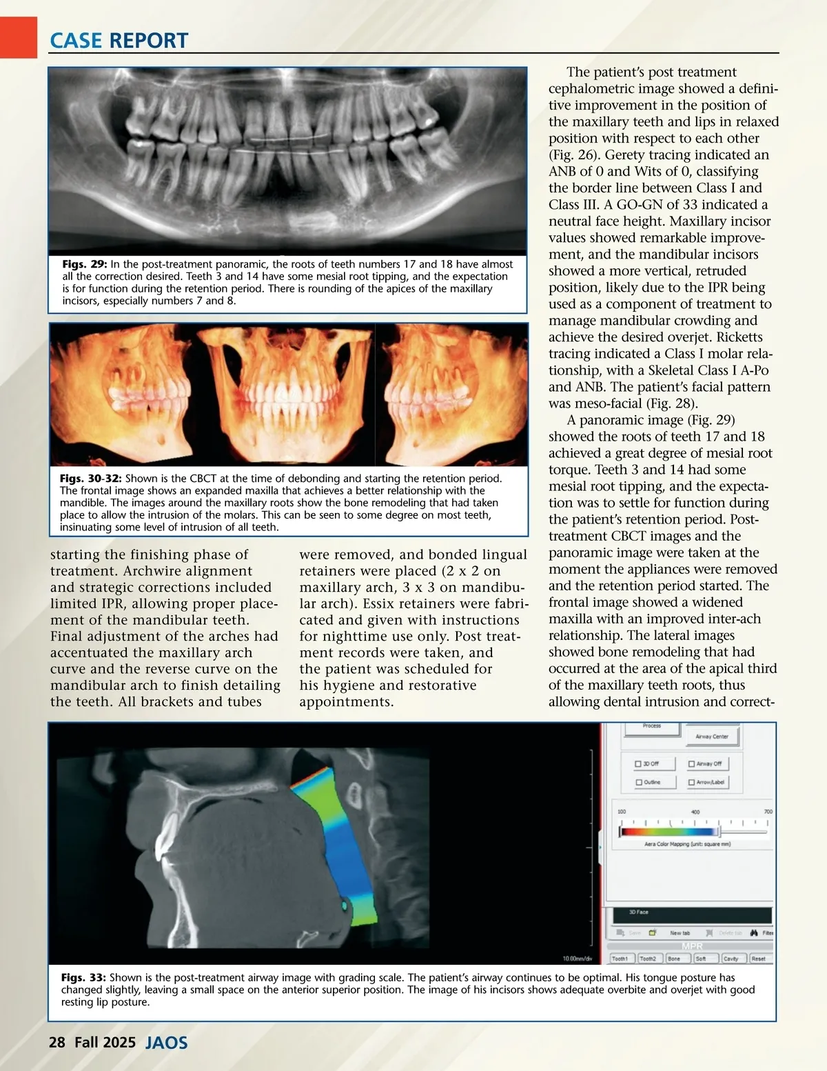
CASE REPORT The patient’s post treatment cephalometric image showed a defini-tive improvement in the position of the maxillary teeth and lips in relaxed position with respect to each other (Fig. 26). Gerety tracing indicated an ANB of 0 and Wits of 0, classifying the border line between Class I and Class III. A GO-GN of 33 indicated a neutral face height. Maxillary incisor values showed remarkable improve-ment, and the mandibular incisors showed a more vertical, retruded position, likely due to the IPR being used as a component of treatment to manage mandibular crowding and achieve the desired overjet. Ricketts tracing indicated a Class I molar rela-tionship, with a Skeletal Class I A-Po and ANB. The patient’s facial pattern was meso-facial (Fig. 28). A panoramic image (Fig. 29) showed the roots of teeth 17 and 18 achieved a great degree of mesial root torque. Teeth 3 and 14 had some mesial root tipping, and the expecta-tion was to settle for function during the patient’s retention period. Post-treatment CBCT images and the panoramic image were taken at the moment the appliances were removed and the retention period started. The frontal image showed a widened maxilla with an improved inter-ach relationship. The lateral images showed bone remodeling that had occurred at the area of the apical third of the maxillary teeth roots, thus allowing dental intrusion and correct-Figs. 29: In the post-treatment panoramic, the roots of teeth numbers 17 and 18 have almost all the correction desired. Teeth 3 and 14 have some mesial root tipping, and the expectation is for function during the retention period. There is rounding of the apices of the maxillary incisors, especially numbers 7 and 8. Figs. 30-32: Shown is the CBCT at the time of debonding and starting the retention period. The frontal image shows an expanded maxilla that achieves a better relationship with the mandible. The images around the maxillary roots show the bone remodeling that had taken place to allow the intrusion of the molars. This can be seen to some degree on most teeth, insinuating some level of intrusion of all teeth. starting the finishing phase of treatment. Archwire alignment and strategic corrections included limited IPR, allowing proper place-ment of the mandibular teeth. Final adjustment of the arches had accentuated the maxillary arch curve and the reverse curve on the mandibular arch to finish detailing the teeth. All brackets and tubes were removed, and bonded lingual retainers were placed (2 x 2 on maxillary arch, 3 x 3 on mandibu-lar arch). Essix retainers were fabri-cated and given with instructions for nighttime use only. Post treat-ment records were taken, and the patient was scheduled for his hygiene and restorative appointments. Figs. 33: Shown is the post-treatment airway image with grading scale. The patient’s airway continues to be optimal. His tongue posture has changed slightly, leaving a small space on the anterior superior position. The image of his incisors shows adequate overbite and overjet with good resting lip posture. 28 Fall 2025 JAOS
Journal of the American Orthodontic Society Fall 2025: Page 28