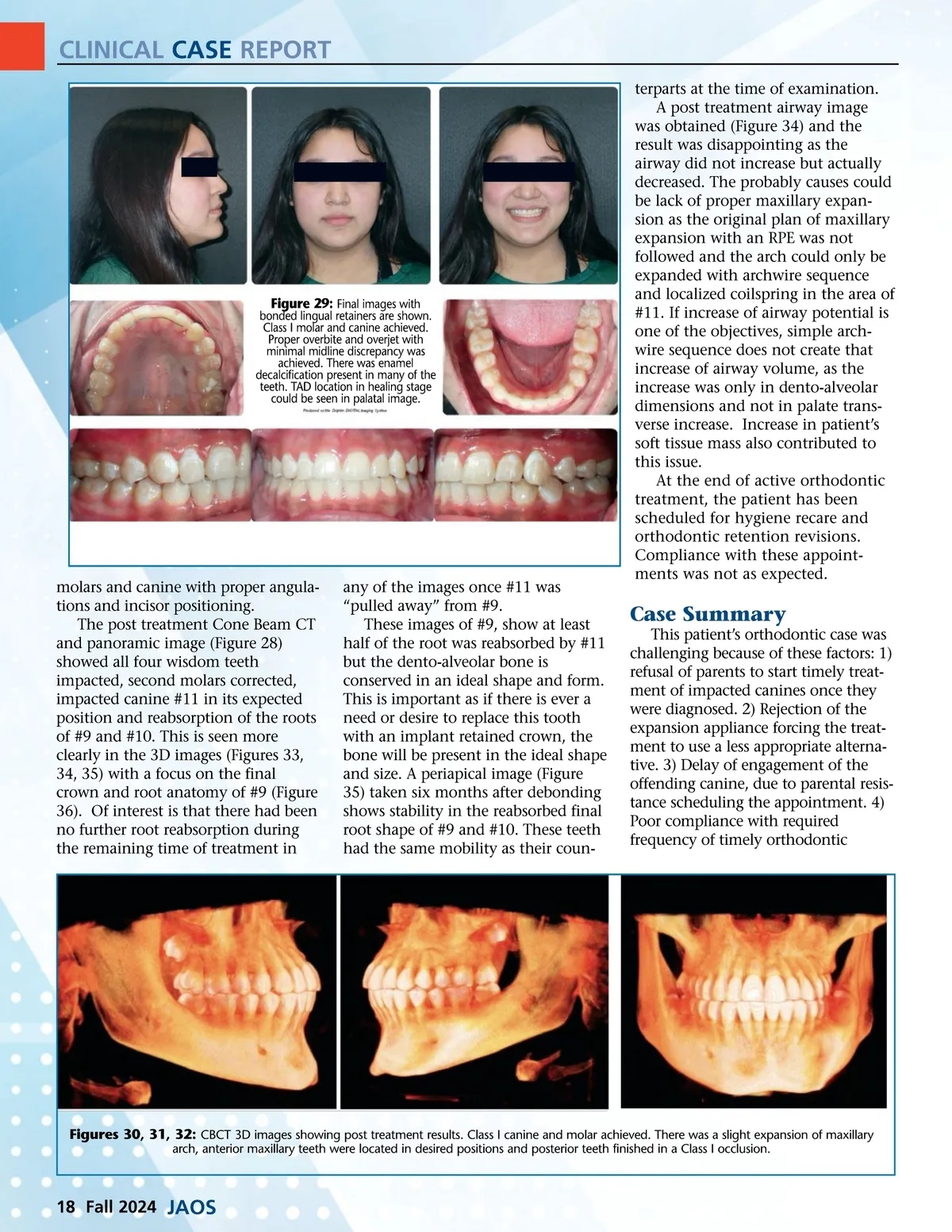
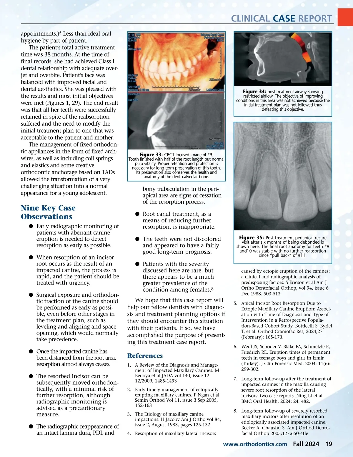
CLINICAL CASE REPORT appointments.) 5 Less than ideal oral hygiene by part of patient. The patient’s total active treatment time was 38 months. At the time of final records, she had achieved Class I dental relationship with adequate over-jet and overbite. Patient’s face was balanced with improved facial and dental aesthetics. She was pleased with the results and most initial objectives were met (Figures 1, 29). The end result was that all her teeth were successfully retained in spite of the reabsorption suffered and the need to modify the initial treatment plan to one that was acceptable to the patient and mother. The management of fixed orthodon-tic appliances in the form of fixed arch-wires, as well as including coil springs and elastics and some creative orthodontic anchorage based on TADs allowed the transformation of a very challenging situation into a normal appearance for a young adolescent. restricted airflow. The objective of improving conditions in this area was not achieved because the initial treatment plan was not followed thus defeating this objective. Figure 34: post treatment airway showing Figure 33: CBCT focused image of #9. Tooth finished with half of the root length but normal pulp vitality. Proper retention and protection is necessary for long term preservation of this tooth. Its preservation also conserves the health and anatomy of the dento-alveolar bone. Nine Key Case Observations b Early radiographic monitoring of patients with aberrant canine eruption is needed to detect resorption as early as possible. b When resorption of an incisor root occurs as the result of an impacted canine, the process is rapid, and the patient should be treated with urgency. b Surgical exposure and orthodon-tic traction of the canine should be performed as early as possi-ble, even before other stages in the treatment plan, such as leveling and aligning and space opening, which would normally take precedence. b Once the impacted canine has been distanced from the root area, resorption almost always ceases. b The resorbed incisor can be subsequently moved orthodon-tically, with a minimal risk of further resorption, although radiographic monitoring is advised as a precautionary measure. b The radiographic reappearance of an intact lamina dura, PDL and bony trabeculation in the peri-apical area are signs of cessation of the resorption process. b Root canal treatment, as a means of reducing further resorption, is inappropriate. b The teeth were not discolored and appeared to have a fairly good long-term prognosis. b Patients with the severity discussed here are rare, but there appears to be a much greater prevalence of the condition among females. 8 We hope that this case report will help our fellow dentists with diagno-sis and treatment planning options if they should encounter this situation with their patients. If so, we have accomplished the purpose of present-ing this treatment case report. visit after six months of being debonded is shown here. The final root anatomy for teeth #9 and10 was stable with no further reabsortion since “pull back” of #11. caused by ectopic eruption of the canines: a clinical and radiographic analysis of predisposing factors. S Ericson et al Am J Ortho Dentofacial Orthop, vol 94, issue 6 Dec 1988. 503-513 5. Apical Incisor Root Resorption Due to Ectopic Maxillary Canine Eruption: Associ-ation with Time of Diagnosis and Type of Intervention in a Retrospective Popula-tion-Based Cohort Study. Botticelli S, Byriel T, et al: Orthod Craniofac Res; 2024;27 (February): 165-173. 6. Wedl JS, Schoder V, Blake FA, Schmelzle R, Friedrich RE. Eruption times of permanent teeth in teenage boys and girls in Izmir (Turkey). J Clin Forensic Med. 2004; 11(6): 299-302. 7. Long-term follow-up after the treatment of impacted canines in the maxilla causing severe root resorption of the lateral incisors: two case reports. Ning LI et al BMC Oral Health. 2024; 24: 482. 8. Long-term follow-up of severely resorbed maxillary incisors after resolution of an etiologically associated impacted canine. Becker A, Chaushu S. Am J Orthod Dento-facial Orthop 2005;127:650-4tle Figure 35: Post treatment periapical recare References 1. A Review of the Diagnosis and Manage-ment of Impacted Maxillary Canines. M Bedoya et al JADA vol 140, issue 12 12/2009, 1485-1493 2. Early timely management of ectopically erupting maxillary canines. P Ngan et al. Semin Orthod Vol 11, issue 3 Sep 2005, 152-163 3. The Etiology of maxillary canine impactions. H Jacoby Am J Ortho vol 84, issue 2, August 1983, pages 125-132 4. Resorption of maxillary lateral incisors www.orthodontics.com Fall 2024 19
Journal of the American Orthodontic Society Fall 2024: Page 19