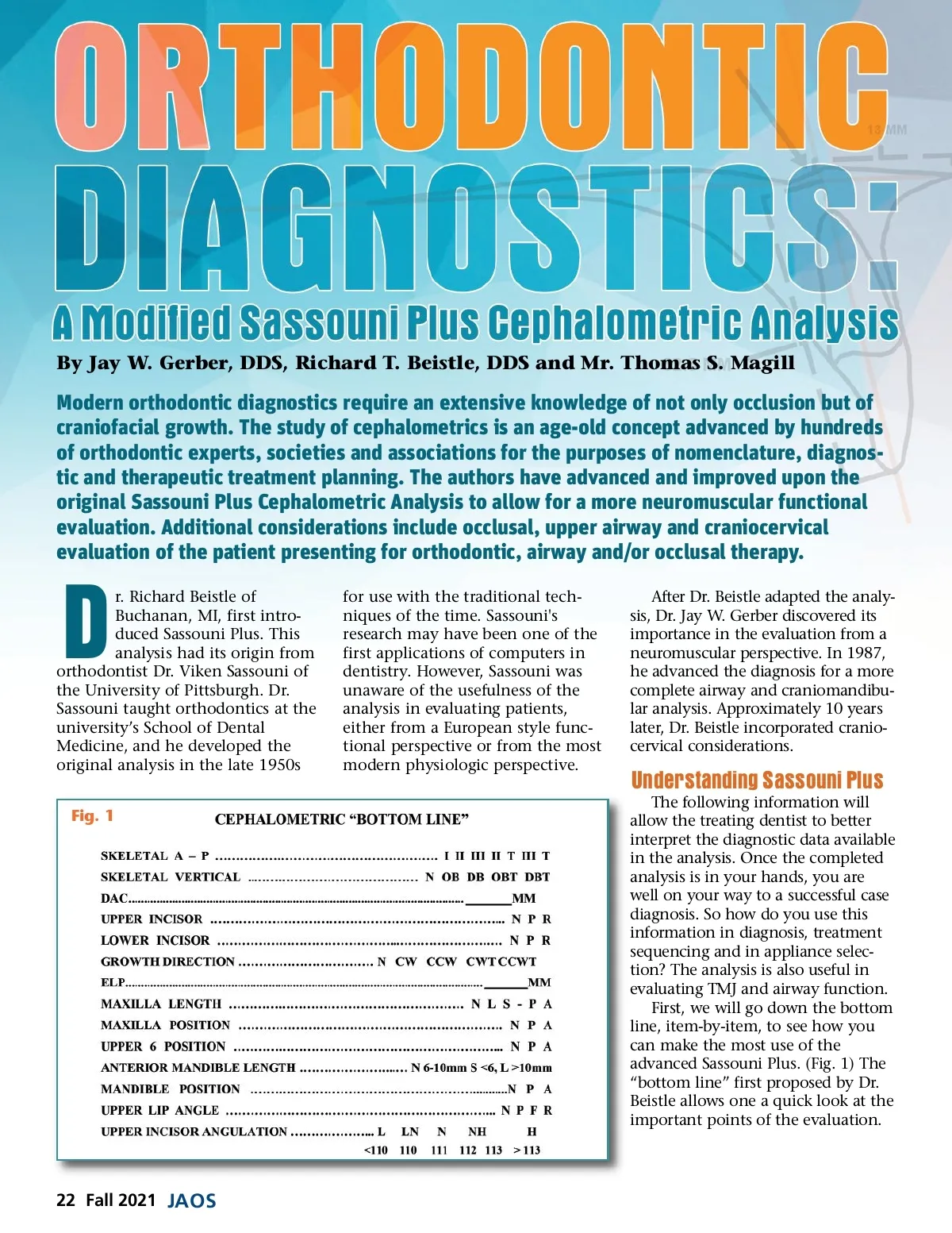
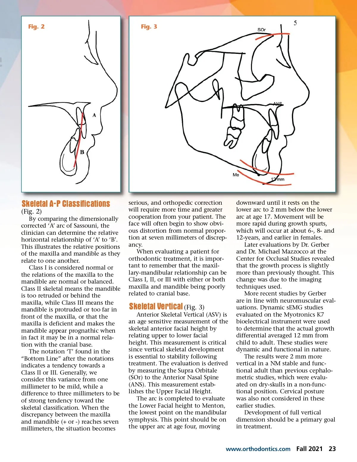
Fig. 2 Fig. 3 Skeletal A-P Classifications (Fig. 2) By comparing the dimensionally corrected ‘A’ arc of Sassouni, the clinician can determine the relative horizontal relationship of ‘A’ to ‘B’. This illustrates the relative positions of the maxilla and mandible as they relate to one another. Class I is considered normal or the relations of the maxilla to the mandible are normal or balanced. Class II skeletal means the mandible is too retruded or behind the maxilla, while Class III means the mandible is protruded or too far in front of the maxilla, or that the maxilla is deficient and makes the mandible appear prognathic when in fact it may be in a normal rela-tion with the cranial base. The notation ‘T’ found in the “Bottom Line” after the notations indicates a tendency towards a Class II or III. Generally, we consider this variance from one millimeter to be mild, while a difference to three millimeters to be of strong tendency toward the skeletal classification. When the discrepancy between the maxilla and mandible (+ or -) reaches seven millimeters, the situation becomes serious, and orthopedic correction will require more time and greater cooperation from your patient. The face will often begin to show obvi-ous distortion from normal propor-tion at seven millimeters of discrep-ancy. When evaluating a patient for orthodontic treatment, it is impor-tant to remember that the maxil-lary-mandibular relationship can be Class I, II, or III with either or both maxilla and mandible being poorly related to cranial base. Skeletal Vertical (Fig. 3) Anterior Skeletal Vertical (ASV) is an age sensitive measurement of the skeletal anterior facial height by relating upper to lower facial height. This measurement is critical since vertical skeletal development is essential to stability following treatment. The evaluation is derived by measuring the Supra Orbitale (SOr) to the Anterior Nasal Spine (ANS). This measurement estab-lishes the Upper Facial Height. The arc is completed to evaluate the Lower Facial height to Menton, the lowest point on the mandibular symphysis. This point should be on the upper arc at age four, moving downward until it rests on the lower arc to 2 mm below the lower arc at age 17. Movement will be more rapid during growth spurts, which will occur at about 6-, 8-and 12-years, and earlier in females. Later evaluations by Dr. Gerber and Dr. Michael Mazzocco at the Center for Occlusal Studies revealed that the growth process is slightly more than previously thought. This change was due to the imaging techniques used. More recent studies by Gerber are in line with neuromuscular eval-uations. Dynamic sEMG studies evaluated on the Myotronics K7 bioelectrical instrument were used to determine that the actual growth differential averaged 12 mm from child to adult. These studies were dynamic and functional in nature. The results were 2 mm more vertical in a NM stable and func-tional adult than previous cephalo-metric studies, which were evalu-ated on dry-skulls in a non-func-tional position. Cervical posture was also not considered in these earlier studies. Development of full vertical dimension should be a primary goal in treatment. www.orthodontics.com Fall 2021 23
Journal of the American Orthodontic Society Fall 2021: Page 23