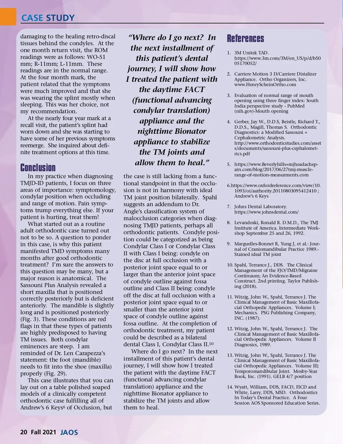
CASE STUDY damaging to the healing retro-discal tissues behind the condyles. At the one month return visit, the ROM readings were as follows: WO-51 mm; R-11mm; L-11mm. These readings are in the normal range. At the four month mark, the patient related that the symptoms were much improved and that she was wearing the splint mostly when sleeping. This was her choice, not my recommendation. At the nearly four year mark at a recall visit, the patient’s splint had worn down and she was starting to have some of her previous symptoms reemerge. She inquired about defi-nite treatment options at this time. Conclusion In my practice when diagnosing TMJD-ID patients, I focus on three areas of importance: symptomology, condylar position when occluding and range of motion. Pain symp-toms trump everything else. If your patient is hurting, treat them! What started out as a routine adult orthodontic case turned out not to be so. A question to ponder in this case, is why this patient manifested TMD symptoms many months after good orthodontic treatment? I’m sure the answers to this question may be many, but a major reason is anatomical. The Sassouni Plus Analysis revealed a short maxilla that is positioned correctly posteriorly but is deficient anteriorly. The mandible is slightly long and is positioned posteriorly (Fig. 3). These conditions are red flags in that these types of patients are highly predisposed to having TM issues. Both condylar eminences are steep. I am reminded of Dr. Len Carapezza’s statement: the foot (mandible) needs to fit into the shoe (maxilla) properly (Fig. 29). This case illustrates that you can lay out on a table polished soaped models of a clinically competent orthodontic case fulfilling all of Andrew’s 6 Keys 6 of Occlusion, but “Where do I go next? In the next installment of this patient’s dental journey, I will show how I treated the patient with the daytime FACT (functional advancing condylar translation) appliance and the nighttime Bionator appliance to stabilize the TM joints and allow them to heal.” the case is still lacking from a func-tional standpoint in that the occlu-sion is not in harmony with ideal TM joint position bilaterally. Spahl suggests an addendum to Dr. Angle’s classification system of malocclusion categories when diag-nosing TMJD patients, perhaps all orthodontic patients. Condyle posi-tion could be categorized as being Condylar Class I or Condylar Class II with Class I being: condyle on the disc at full occlusion with a posterior joint space equal to or larger than the anterior joint space of condyle outline against fossa outline and Class II being: condyle off the disc at full occlusion with a posterior joint space equal to or smaller than the anterior joint space of condyle outline against fossa outline. At the completion of orthodontic treatment, my patient could be described as a bilateral dental Class I, Condylar Class II. 10 Where do I go next? In the next installment of this patient’s dental journey, I will show how I treated the patient with the daytime FACT (functional advancing condylar translation) appliance and the nighttime Bionator appliance to stabilize the TM joints and allow them to heal. References 1. 3M Unitek TAD. https://www.3m.com/3M/en_US/p/d/b50 05170052/ 2. Carriere Motion 3 D/Carriere Distalizer Appliance. Ortho Organizers, Inc. www.HenryScheinOrtho.com 3. Evaluation of normal range of mouth opening using three finger index: South India perspective study -PubMed (nih.gov)-Mouth opening 4. Gerber, Jay W., D.D.S, Beistle, Richard T., D.D.S., Magill, Thomas S. Orthodontic Diagnostics: a Modified Sassouni + Cephalometric Analysis. http://www.orthodonticstudies.com/asset s/documents/sassouni-plus-cephalomet-rics.pdf 5. https://www.Beverlyhills+mjheadachep-ain.com/blog/2017/06/27tmj-muscle-range-of-motion-measurments.com 6.https://www.oxfordreference.com/view/10. 1093/oi/authority.20110803095412410 ; Andrew’s 6 Keys 7. Johns Dental Laboratory. https://www.johnsdental.com/ 8. Levandoski, Ronald R. D.M.D., The TMJ Institute of America. Intermediate Work-shop September 25 and 26, 1992. 9. Marguelles-Bonnet R, Yung J, et al.: Jour-nal of Craniomandibular Practice 1989.-Stained ideal TM joint 10. Spahl, Terrance J., DDS. The Clinical Management of the FJO/TMD/Migraine Continuum; An Evidence-Based Construct. 2nd printing. Taylor Publish-ing (2018). 11. Witzig, John W., Spahl, Terrance J. The Clinical Management of Basic Maxillofa-cial Orthopedic Appliances. Volume I; Mechanics. PSG Publishing Company, INC. (1987). 12. Witzig, John W., Spahl, Terrance J. The Clinical Management of Basic Maxillofa-cial Orthopedic Appliances. Volume II Diagnosics, 1989. 13. Witzig, John W., Spahl, Terrance J. The Clinical Management of Basic Maxillofa-cial Orthopedic Appliances. Volume III; Temporomandibular Joint. Mosby-Year Book, Inc. (1991). GELB 4/7 position 14. Wyatt, William, DDS, FACD, FICD and White, Larry, DDS, MSD. Orthodontics In Today’s Dental Practice. A Four Session AOS Sponsored Education Series. 20 Fall 2021 JAOS
Journal of the American Orthodontic Society Fall 2021: Page 20