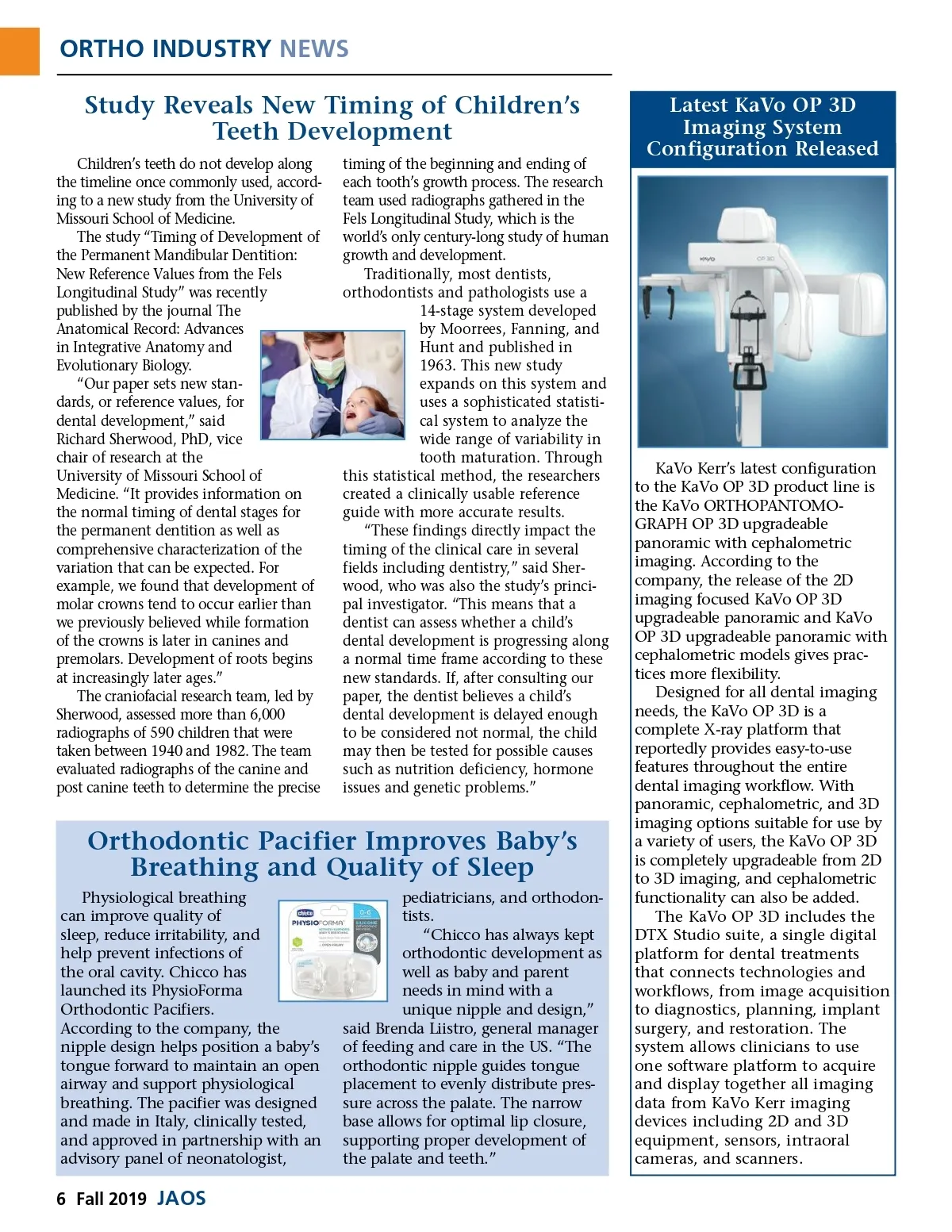
ORTHO INDUSTRY NEWS Study Reveals New Timing of Children’s Teeth Development Children’s teeth do not develop along the timeline once commonly used, accord-ing to a new study from the University of Missouri School of Medicine. The study “Timing of Development of the Permanent Mandibular Dentition: New Reference Values from the Fels Longitudinal Study” was recently published by the journal The Anatomical Record: Advances in Integrative Anatomy and Evolutionary Biology. “Our paper sets new stan-dards, or reference values, for dental development,” said Richard Sherwood, PhD, vice chair of research at the University of Missouri School of Medicine. “It provides information on the normal timing of dental stages for the permanent dentition as well as comprehensive characterization of the variation that can be expected. For example, we found that development of molar crowns tend to occur earlier than we previously believed while formation of the crowns is later in canines and premolars. Development of roots begins at increasingly later ages.” The craniofacial research team, led by Sherwood, assessed more than 6,000 radiographs of 590 children that were taken between 1940 and 1982. The team evaluated radiographs of the canine and post canine teeth to determine the precise timing of the beginning and ending of each tooth’s growth process. The research team used radiographs gathered in the Fels Longitudinal Study, which is the world’s only century-long study of human growth and development. Traditionally, most dentists, orthodontists and pathologists use a 14-stage system developed by Moorrees, Fanning, and Hunt and published in 1963. This new study expands on this system and uses a sophisticated statisti-cal system to analyze the wide range of variability in tooth maturation. Through this statistical method, the researchers created a clinically usable reference guide with more accurate results. “These findings directly impact the timing of the clinical care in several fields including dentistry,” said Sher-wood, who was also the study’s princi-pal investigator. “This means that a dentist can assess whether a child’s dental development is progressing along a normal time frame according to these new standards. If, after consulting our paper, the dentist believes a child’s dental development is delayed enough to be considered not normal, the child may then be tested for possible causes such as nutrition deficiency, hormone issues and genetic problems.” Latest KaVo OP 3D Imaging System Configuration Released Orthodontic Pacifier Improves Baby’s Breathing and Quality of Sleep Physiological breathing can improve quality of sleep, reduce irritability, and help prevent infections of the oral cavity. Chicco has launched its PhysioForma Orthodontic Pacifiers. According to the company, the nipple design helps position a baby’s tongue forward to maintain an open airway and support physiological breathing. The pacifier was designed and made in Italy, clinically tested, and approved in partnership with an advisory panel of neonatologist, pediatricians, and orthodon-tists. “Chicco has always kept orthodontic development as well as baby and parent needs in mind with a unique nipple and design,” said Brenda Liistro, general manager of feeding and care in the US. “The orthodontic nipple guides tongue placement to evenly distribute pres-sure across the palate. The narrow base allows for optimal lip closure, supporting proper development of the palate and teeth.” KaVo Kerr’s latest configuration to the KaVo OP 3D product line is the KaVo ORTHOPANTOMO-GRAPH OP 3D upgradeable panoramic with cephalometric imaging. According to the company, the release of the 2D imaging focused KaVo OP 3D upgradeable panoramic and KaVo OP 3D upgradeable panoramic with cephalometric models gives prac-tices more flexibility. Designed for all dental imaging needs, the KaVo OP 3D is a complete X-ray platform that reportedly provides easy-to-use features throughout the entire dental imaging workflow. With panoramic, cephalometric, and 3D imaging options suitable for use by a variety of users, the KaVo OP 3D is completely upgradeable from 2D to 3D imaging, and cephalometric functionality can also be added. The KaVo OP 3D includes the DTX Studio suite, a single digital platform for dental treatments that connects technologies and workflows, from image acquisition to diagnostics, planning, implant surgery, and restoration. The system allows clinicians to use one software platform to acquire and display together all imaging data from KaVo Kerr imaging devices including 2D and 3D equipment, sensors, intraoral cameras, and scanners. 6 Fall 2019 JAOS
Journal of the American Orthodontic Society Fall 2019: Page 6