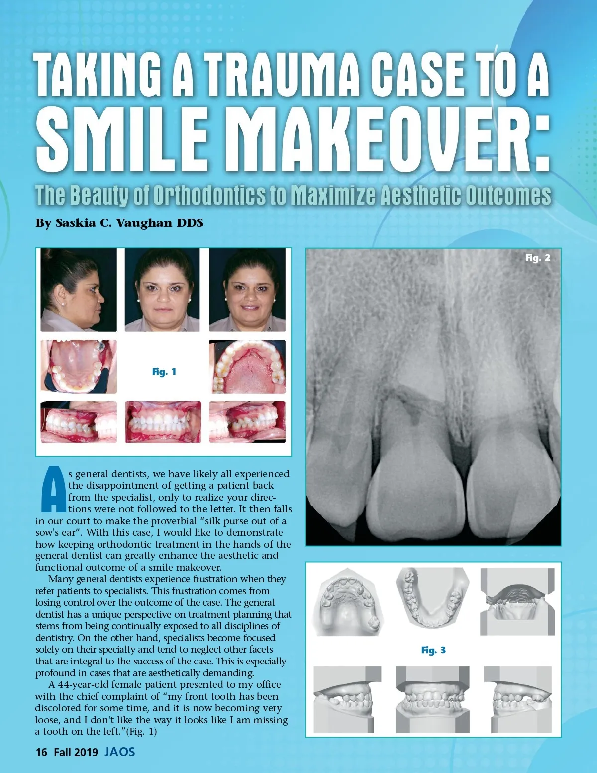
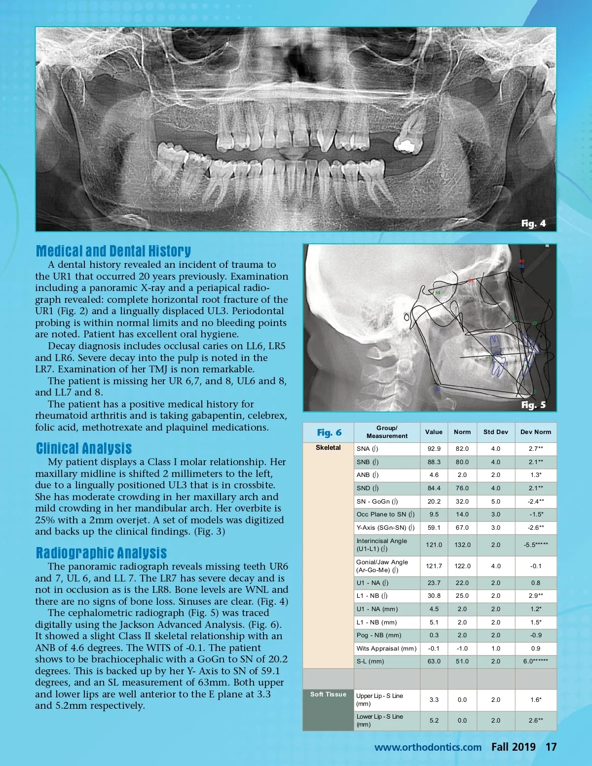
Fig. 4 Medical and Dental History A dental history revealed an incident of trauma to the UR1 that occurred 20 years previously. Examination including a panoramic X-ray and a periapical radio-graph revealed: complete horizontal root fracture of the UR1 (Fig. 2) and a lingually displaced UL3. Periodontal probing is within normal limits and no bleeding points are noted. Patient has excellent oral hygiene. Decay diagnosis includes occlusal caries on LL6, LR5 and LR6. Severe decay into the pulp is noted in the LR7. Examination of her TMJ is non remarkable. The patient is missing her UR 6,7, and 8, UL6 and 8, and LL7 and 8. The patient has a positive medical history for rheumatoid arthritis and is taking gabapentin, celebrex, folic acid, methotrexate and plaquinel medications. Fig. 5 Fig. 6 Group/ Measurement Value 92.9 88.3 84.4 20.2 59.1 9.5 4.6 Norm 82.0 80.0 76.0 32.0 14.0 67.0 2.0 Std Dev 4.0 4.0 2.0 4.0 5.0 3.0 3.0 Dev Norm 2.7** 2.1** 2.1** 1.3* Clinical Analysis My patient displays a Class I molar relationship. Her maxillary midline is shifted 2 millimeters to the left, due to a lingually positioned UL3 that is in crossbite. She has moderate crowding in her maxillary arch and mild crowding in her mandibular arch. Her overbite is 25% with a 2mm overjet. A set of models was digitized and backs up the clinical findings. (Fig. 3) Skeletal SNA (∫) SNB (∫) ANB (∫) SND (∫) SN -GoGn (∫) Occ Plane to SN (∫) -2.4** -2.6** -1.5* Y-Axis (SGn-SN) (∫) Radiographic Analysis The panoramic radiograph reveals missing teeth UR6 and 7, UL 6, and LL 7. The LR7 has severe decay and is not in occlusion as is the LR8. Bone levels are WNL and there are no signs of bone loss. Sinuses are clear. (Fig. 4) The cephalometric radiograph (Fig. 5) was traced digitally using the Jackson Advanced Analysis. (Fig. 6). It showed a slight Class II skeletal relationship with an ANB of 4.6 degrees. The WITS of -0.1. The patient shows to be brachiocephalic with a GoGn to SN of 20.2 degrees. This is backed up by her Y-Axis to SN of 59.1 degrees, and an SL measurement of 63mm. Both upper and lower lips are well anterior to the E plane at 3.3 and 5.2mm respectively. Gonial/Jaw Angle (Ar-Go-Me) (∫) U1 -NA (∫) L1 -NB (∫) Interincisal Angle (U1-L1) (∫) 121.0 121.7 23.7 30.8 4.5 5.1 -0.1 0.3 132.0 122.0 22.0 25.0 2.0 2.0 -1.0 2.0 2.0 -5.5***** -0.1 2.9** 1.2* 1.5* -0.9 0.9 0.8 2.0 2.0 2.0 2.0 2.0 1.0 2.0 4.0 U1 -NA (mm) L1 -NB (mm) Pog -NB (mm) S-L (mm) Wits Appraisal (mm) 63.0 51.0 6.0****** Soft Tissue Upper Lip -S Line (mm) Lower Lip -S Line (mm) 3.3 0.0 2.0 1.6* 5.2 0.0 2.0 2.6** www.orthodontics.com Fall 2019 17
Journal of the American Orthodontic Society Fall 2019: Page 17