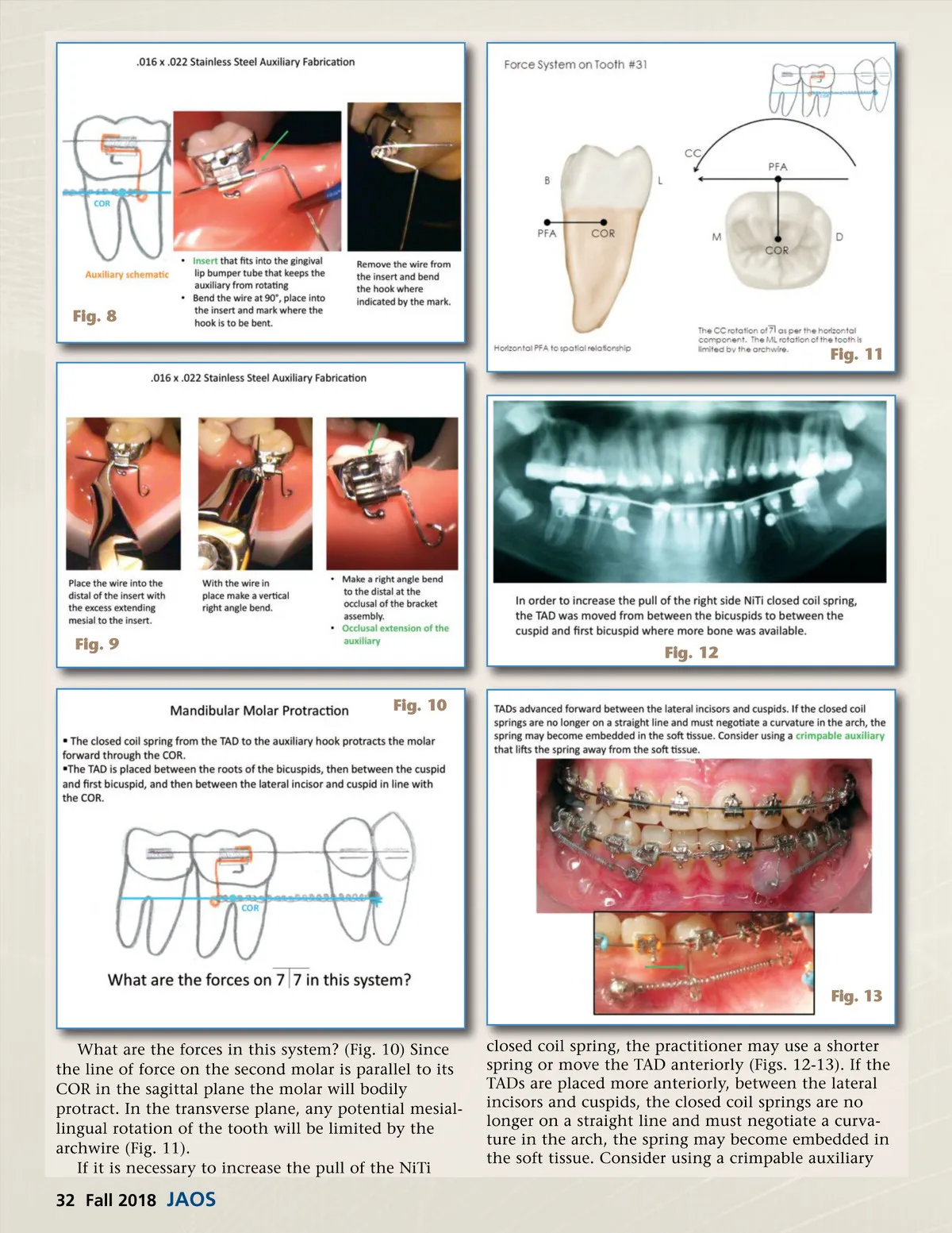
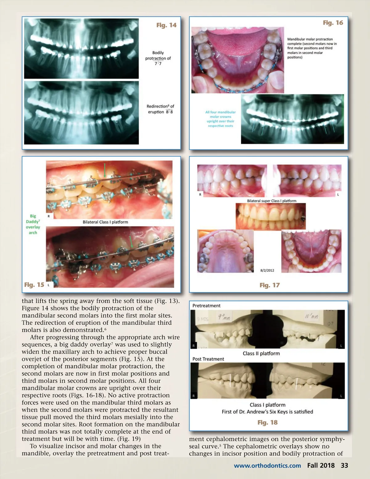
Fig. 14 Fig. 16 Fig. 15 that lifts the spring away from the soft tissue (Fig. 13). Figure 14 shows the bodily protraction of the mandibular second molars into the first molar sites. The redirection of eruption of the mandibular third molars is also demonstrated. 6 After progressing through the appropriate arch wire sequences, a big daddy overlay 7 was used to slightly widen the maxillary arch to achieve proper buccal overjet of the posterior segments (Fig. 15). At the completion of mandibular molar protraction, the second molars are now in first molar positions and third molars in second molar positions. All four mandibular molar crowns are upright over their respective roots (Figs. 16-18). No active protraction forces were used on the mandibular third molars as when the second molars were protracted the resultant tissue pull moved the third molars mesially into the second molar sites. Root formation on the mandibular third molars was not totally complete at the end of treatment but will be with time. (Fig. 19) To visualize incisor and molar changes in the mandible, overlay the pretreatment and post treat-Fig. 17 Fig. 18 ment cephalometric images on the posterior symphy-seal curve. 5 The cephalometric overlays show no changes in incisor position and bodily protraction of www.orthodontics.com Fall 2018 33
Journal of the American Orthodontic Society Fall 2018: Page 33