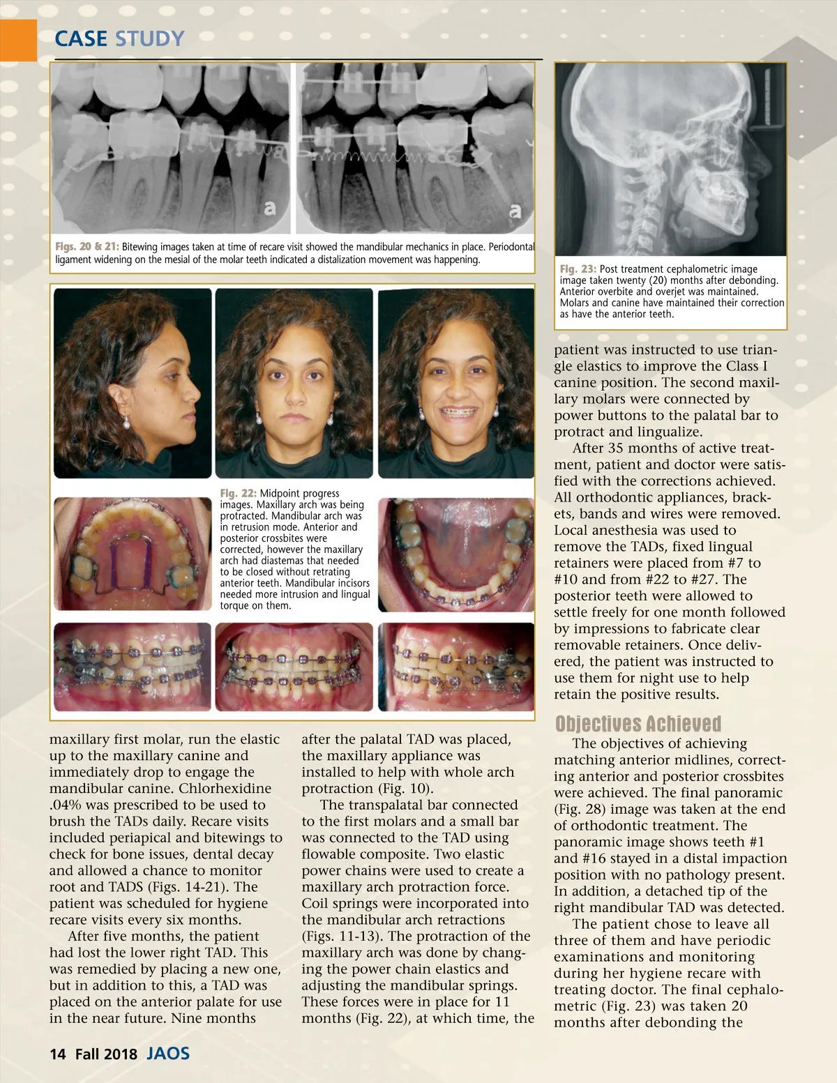
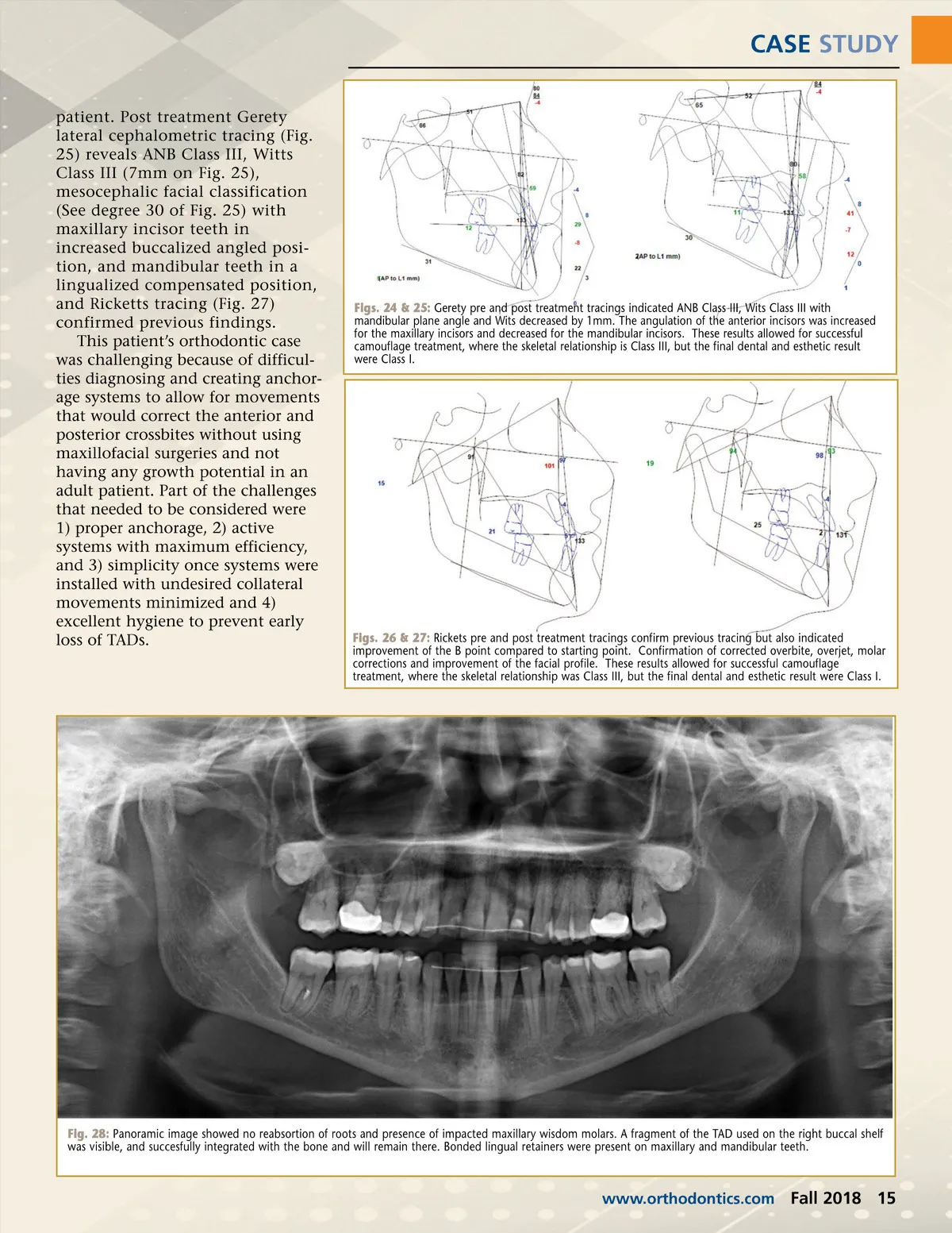
CASE STUDY patient. Post treatment Gerety lateral cephalometric tracing (Fig. 25) reveals ANB Class III, Witts Class III (7mm on Fig. 25), mesocephalic facial classification (See degree 30 of Fig. 25) with maxillary incisor teeth in increased buccalized angled posi-tion, and mandibular teeth in a lingualized compensated position, and Ricketts tracing (Fig. 27) confirmed previous findings. This patient’s orthodontic case was challenging because of difficul-ties diagnosing and creating anchor-age systems to allow for movements that would correct the anterior and posterior crossbites without using maxillofacial surgeries and not having any growth potential in an adult patient. Part of the challenges that needed to be considered were 1) proper anchorage, 2) active systems with maximum efficiency, and 3) simplicity once systems were installed with undesired collateral movements minimized and 4) excellent hygiene to prevent early loss of TADs. Figs. 24 & 25: Gerety pre and post treatment tracings indicated ANB Class III, Wits Class III with mandibular plane angle and Wits decreased by 1mm. The angulation of the anterior incisors was increased for the maxillary incisors and decreased for the mandibular incisors. These results allowed for successful camouflage treatment, where the skeletal relationship is Class III, but the final dental and esthetic result were Class I. Figs. 26 & 27: Rickets pre and post treatment tracings confirm previous tracing but also indicated improvement of the B point compared to starting point. Confirmation of corrected overbite, overjet, molar corrections and improvement of the facial profile. These results allowed for successful camouflage treatment, where the skeletal relationship was Class III, but the final dental and esthetic result were Class I. Fig. 28: Panoramic image showed no reabsortion of roots and presence of impacted maxillary wisdom molars. A fragment of the TAD used on the right buccal shelf was visible, and succesfully integrated with the bone and will remain there. Bonded lingual retainers were present on maxillary and mandibular teeth. www.orthodontics.com Fall 2018 15
Journal of the American Orthodontic Society Fall 2018: Page 15