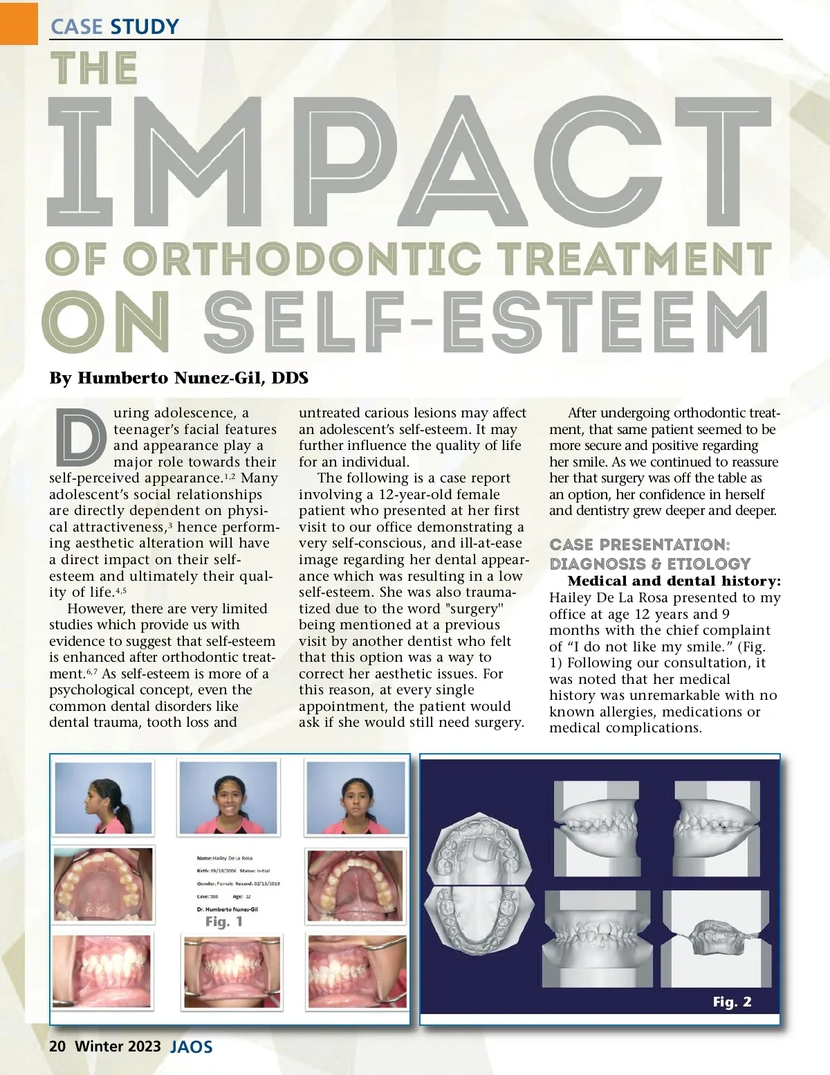
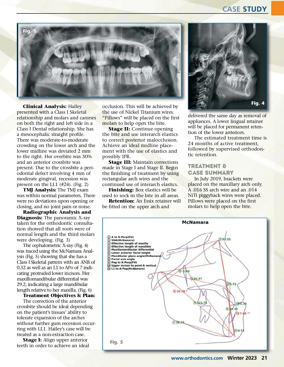
CASE STUDY Fig. 3 Clinical Analysis: Hailey presented with a Class I Skeletal relationship and molars and canines on both the right and left side in a Class I Dental relationship. She has a mesocephalic straight profile. There was moderate-to-moderate crowding on the lower arch and the lower midline was deviated 2 mm to the right. Her overbite was 30% and an anterior crossbite was present. Due to the crossbite a peri-odontal defect involving 4 mm of moderate gingival, recession was present on the LL1 (#24). (Fig. 2) TMJ Analysis: The TMJ exam was within normal parameters. There were no deviations upon opening or closing, and no joint pain or noise. Radiographic Analysis and Diagnosis: The panoramic X-ray taken for the orthodontic consulta-tion showed that all roots were of normal length and the third molars were developing. (Fig. 3) The cephalometric X-ray (Fig. 4) was traced using the McNamara Anal-ysis (Fig. 5) showing that she has a Class I Skeletal pattern with an ANB of 0.32 as well as an L1 to APo of 7 indi-cating protruded lower incisors. Her maxillomandibular differential was 29.2, indicating a large mandibular length relative to her maxilla. (Fig. 6) Treatment Objectives & Plan: The correction of the anterior crossbite should be ideal depending on the patient’s tissues’ ability to tolerate expansion of the arches without further gum recession occur-ring with LL1. Hailey’s case will be treated as a non-extraction case. Stage I: Align upper anterior teeth in order to achieve an ideal occlusion. This will be achieved by the use of Nickel Titanium wires. “Pillows” will be placed on the first molars to help open the bite. Stage II: Continue opening the bite and use interarch elastics to correct posterior malocclusion. Achieve an ideal midline place-ment with the use of elastics and possibly IPR. Stage III: Maintain corrections made in Stage I and Stage II. Begin the finishing of treatment by using rectangular arch wires and the continued use of interarch elastics. Finishing: Box elastics will be used to sock in the bite in all areas. Retention: An Essix retainer will be fitted on the upper arch and Fig. 4 delivered the same day as removal of appliances. A lower lingual retainer will be placed for permanent reten-tion of the lower anteriors. The estimated treatment time is 24 months of active treatment, followed by supervised orthodon-tic retention. Treatment & Case Summary In July 2019, brackets were placed on the maxillary arch only. A .016 SS arch wire and an .014 NiTi piggyback wires were placed. Pillows were placed on the first molars to help open the bite. Fig. 5 www.orthodontics.com Winter 2023 21
Journal of the American Orthodontic Society Winter 2023: Page 21