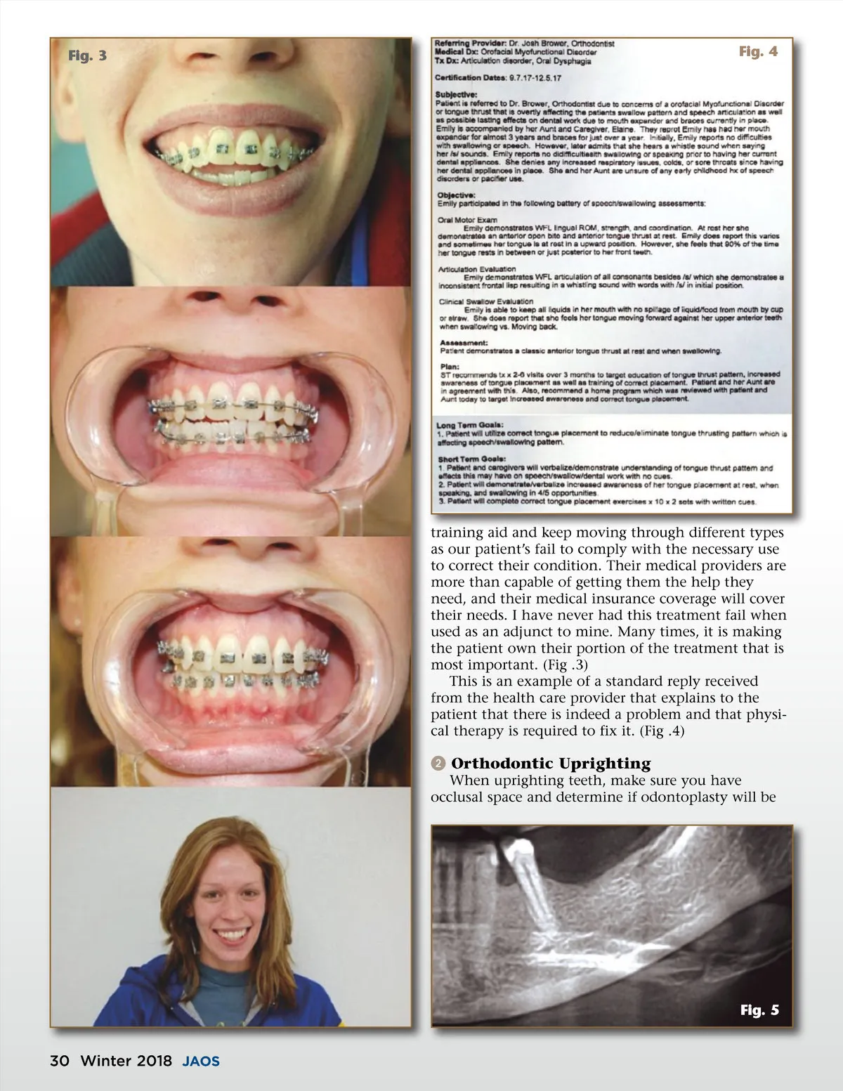
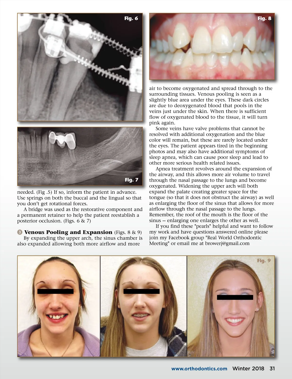
Fig. 6 Fig. 8 Fig. 7 needed. (Fig .5) If so, inform the patient in advance. Use springs on both the buccal and the lingual so that you don’t get rotational forces. A bridge was used as the restorative component and a permanent retainer to help the patient reestablish a posterior occlusion. (Figs. 6 & 7) ᕣ Venous Pooling and Expansion (Figs. 8 & 9) By expanding the upper arch, the sinus chamber is also expanded allowing both more airflow and more air to become oxygenated and spread through to the surrounding tissues. Venous pooling is seen as a slightly blue area under the eyes. These dark circles are due to deoxygenated blood that pools in the veins just under the skin. When there is sufficient flow of oxygenated blood to the tissue, it will turn pink again. Some veins have valve problems that cannot be resolved with additional oxygenation and the blue color will remain, but these are rarely located under the eyes. The patient appears tired in the beginning photos and may also have additional symptoms of sleep apnea, which can cause poor sleep and lead to other more serious health related issues. Apnea treatment revolves around the expansion of the airway, and this allows more air volume to travel through the nasal passage to the lungs and become oxygenated. Widening the upper arch will both expand the palate creating greater space for the tongue (so that it does not obstruct the airway) as well as enlarging the floor of the sinus that allows for more airflow through the nasal passage to the lungs. Remember, the roof of the mouth is the floor of the sinus --enlarging one enlarges the other as well. If you find these "pearls" helpful and want to follow my work and have questions answered online please join my Facebook group "Real World Orthodontic Meeting" or email me at browerj@gmail.com Fig. 9 www.orthodontics.com Winter 2018 31
Journal of the American Orthodontic Society Winter 2018: Page 31