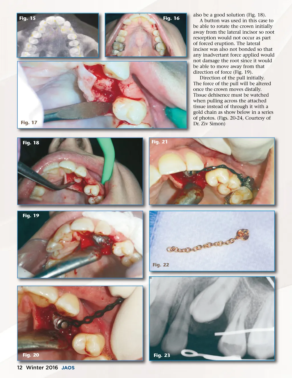
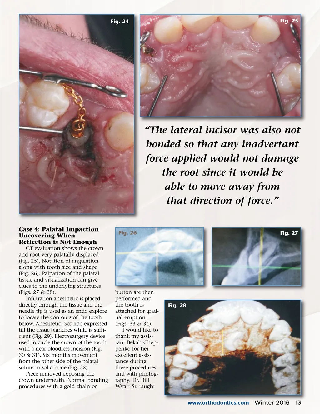
Fig. 24 Fig. 25 “The lateral incisor was also not bonded so that any inadvertant force applied would not damage the root since it would be able to move away from that direction of force.” Case 4: Palatal Impaction Uncovering When Reflection is Not Enough CT evaluation shows the crown and root very palatally displaced (Fig. 25). Notation of angulation along with tooth size and shape (Fig. 26). Palpation of the palatal tissue and visualization can give clues to the underlying structures (Figs. 27 & 28). Infiltration anesthetic is placed directly through the tissue and the needle tip is used as an endo explore to locate the contours of the tooth below. Anesthetic .5cc lido expressed till the tissue blanches white is suffi-cient (Fig. 29). Electrosurgery device used to circle the crown of the tooth with a near bloodless incision (Fig. 30 & 31). Six months movement from the other side of the palatal suture in solid bone (Fig. 32). Piece removed exposing the crown underneath. Normal bonding procedures with a gold chain or Fig. 26 Fig. 27 button are then performed and the tooth is attached for grad-ual eruption (Figs. 33 & 34). I would like to thank my assis-tant Bekah Chep-penko for her excellent assis-tance during these procedures and with photog-raphy. Dr. Bill Wyatt Sr. taught Fig. 28 www.orthodontics.com Winter 2016 13
Journal of the American Orthodontic Society Winter 2016: Page 13