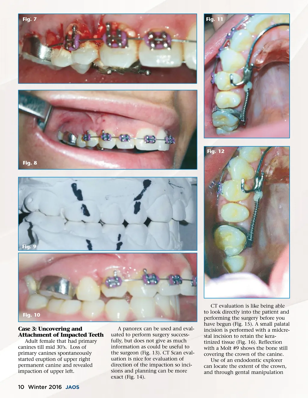
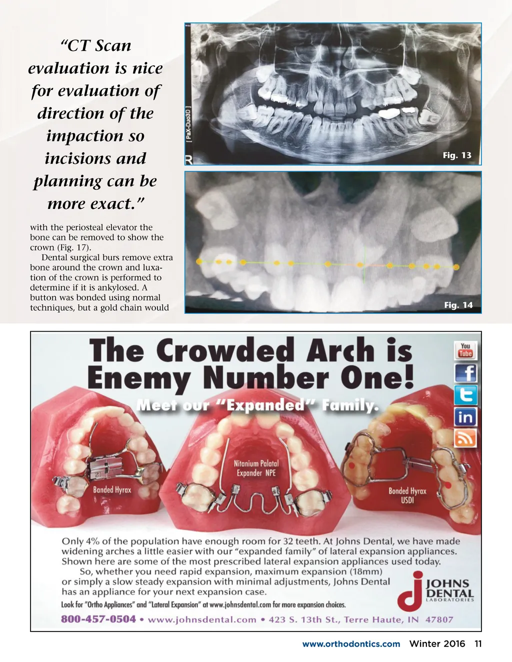
Fig. 7 Fig. 11 Fig. 12 Fig. 8 Fig. 9 Fig. 10 Case 3: Uncovering and Attachment of Impacted Teeth Adult female that had primary canines till mid 30’s. Loss of primary canines spontaneously started eruption of upper right permanent canine and revealed impaction of upper left. A panorex can be used and eval-uated to perform surgery success-fully, but does not give as much information as could be useful to the surgeon (Fig. 13). CT Scan eval-uation is nice for evaluation of direction of the impaction so inci-sions and planning can be more exact (Fig. 14). CT evaluation is like being able to look directly into the patient and performing the surgery before you have begun (Fig. 15). A small palatal incision is performed with a midcre-stal incision to retain the kera-tinized tissue (Fig. 16). Reflection with a Molt #9 shows the bone still covering the crown of the canine. Use of an endodontic explorer can locate the extent of the crown, and through gental manipulation 10 Winter 2016 JAOS
Journal of the American Orthodontic Society Winter 2016: Page 10