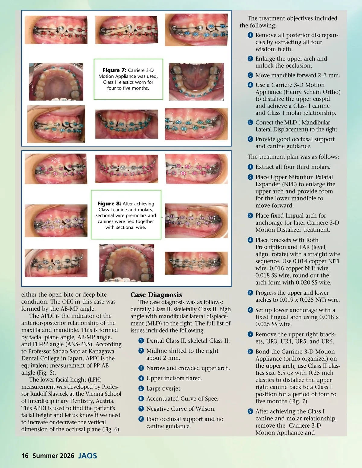
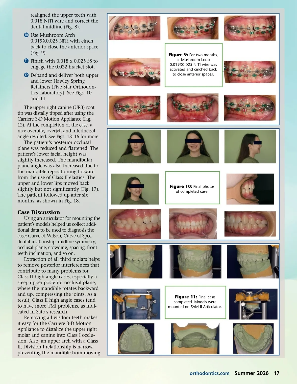
The treatment objectives included the following: ᕡ Remove all posterior discrepan-cies by extracting all four wisdom teeth. ᕢ Enlarge the upper arch and unlock the occlusion. ᕣ Move mandible forward 2–3 mm. ᕤ Use a Carriere 3-D Motion Appliance (Henry Schein Ortho) to distalize the upper cuspid and achieve a Class I canine and Class I molar relationship. ᕥ Correct the MLD ( Mandibular Lateral Displacement) to the right. ᕦ Provide good occlusal support and canine guidance. The treatment plan was as follows: ᕡ Extract all four third molars. ᕢ Place Upper Nitanium Palatal Expander (NPE) to enlarge the upper arch and provide room for the lower mandible to move forward. ᕣ Place fixed lingual arch for anchorage for later Carriere 3-D Motion Distalizer treatment. ᕤ Place brackets with Roth Prescription and LAR (level, align, rotate) with a straight wire sequence. Use 0.014 copper NiTi wire, 0.016 copper NiTi wire, 0.018 SS wire, round out the arch form with 0.020 SS wire. either the open bite or deep bite condition. The ODI in this case was formed by the AB-MP angle. The APDI is the indicator of the anterior-posterior relationship of the maxilla and mandible. This is formed by facial plane angle, AB-MP angle, and FH-PP angle (ANS-PNS). According to Professor Sadao Sato at Kanagawa Dental College in Japan, APDI is the equivalent measurement of PP-AB angle (Fig. 5). The lower facial height (LFH) measurement was developed by Profes-sor Rudolf Slavicek at the Vienna School of Interdisciplinary Dentistry, Austria. This APDI is used to find the patient’s facial height and let us know if we need to increase or decrease the vertical dimension of the occlusal plane (Fig. 6). Figure 7: Carriere 3-D Motion Appliance was used, Class II elastics worn for four to five months. Figure 8: After achieving Class I canine and molars, sectional wire premolars and canines were tied together with sectional wire. Case Diagnosis The case diagnosis was as follows: dentally Class II, skeletally Class II, high angle with mandibular lateral displace-ment (MLD) to the right. The full list of issues included the following: ᕡ Dental Class II, skeletal Class II. ᕢ Midline shifted to the right about 2 mm. ᕣ Narrow and crowded upper arch. ᕤ Upper incisors flared. ᕥ Large overjet. ᕦ Accentuated Curve of Spee. ᕧ Negative Curve of Wilson. ᕨ Poor occlusal support and no canine guidance. ᕥ Progress the upper and lower arches to 0.019 x 0.025 NiTi wire. ᕦ Set up lower anchorage with a fixed lingual arch using 0.018 x 0.025 SS wire. ᕧ Remove the upper right brack-ets, UR3, UR4, UR5, and UR6. ᕨ Bond the Carriere 3-D Motion Appliance (ortho organizer) on the upper arch, use Class II elas-tics size 6.5 oz with 0.25 inch elastics to distalize the upper right canine back to a Class I position for a period of four to five months (Fig. 7). ᕩ After achieving the Class I canine and molar relationship, remove the Carriere 3-D Motion Appliance and 16 Summer 2026 JAOS
Journal of the American Orthodontic Society Summer 2026: Page 16