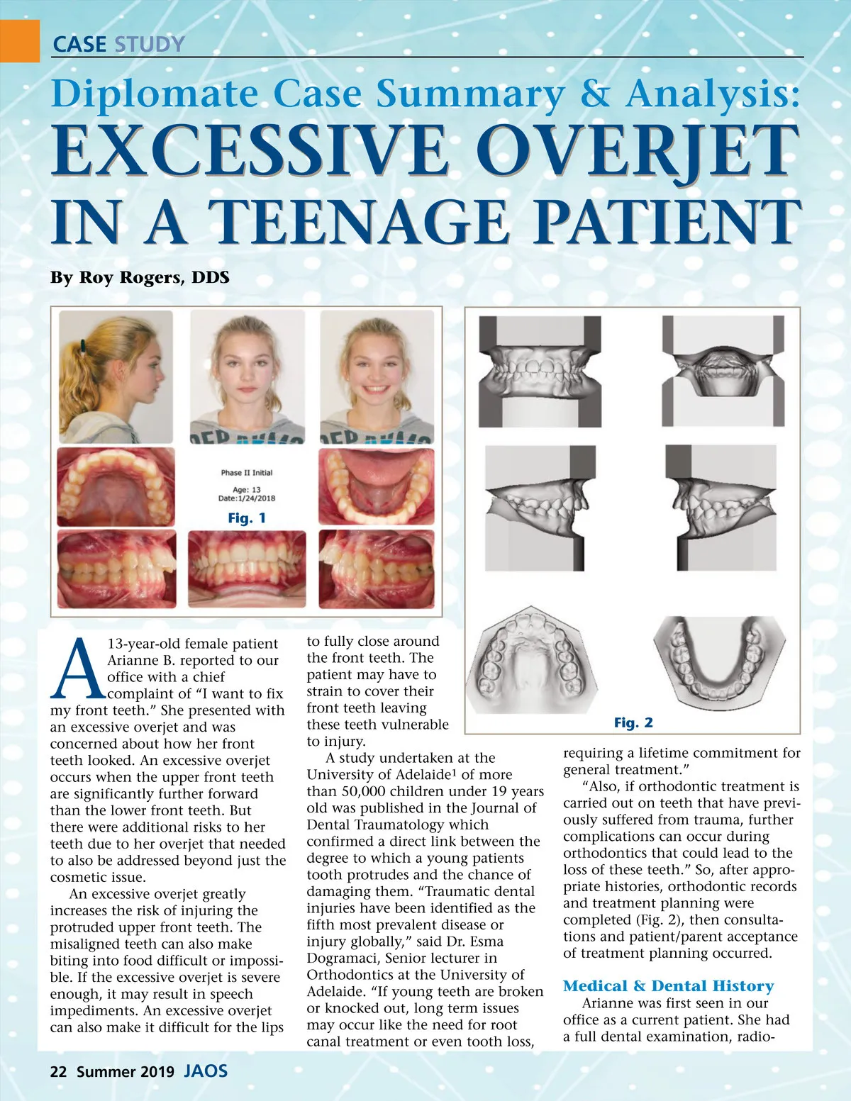
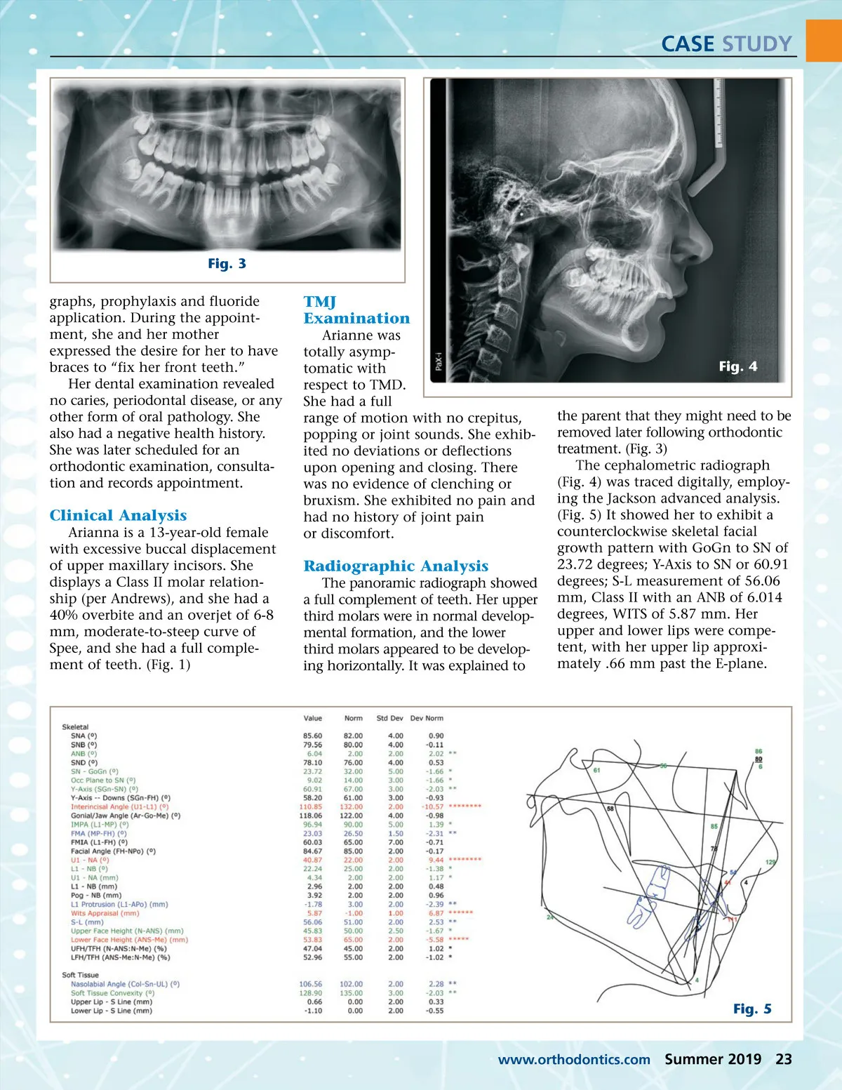
CASE STUDY Fig. 3 graphs, prophylaxis and fluoride application. During the appoint-ment, she and her mother expressed the desire for her to have braces to “fix her front teeth.” Her dental examination revealed no caries, periodontal disease, or any other form of oral pathology. She also had a negative health history. She was later scheduled for an orthodontic examination, consulta-tion and records appointment. TMJ Examination Arianne was totally asymp-tomatic with respect to TMD. She had a full range of motion with no crepitus, popping or joint sounds. She exhib-ited no deviations or deflections upon opening and closing. There was no evidence of clenching or bruxism. She exhibited no pain and had no history of joint pain or discomfort. Fig. 4 Clinical Analysis Arianna is a 13-year-old female with excessive buccal displacement of upper maxillary incisors. She displays a Class II molar relation-ship (per Andrews), and she had a 40% overbite and an overjet of 6-8 mm, moderate-to-steep curve of Spee, and she had a full comple-ment of teeth. (Fig. 1) Radiographic Analysis The panoramic radiograph showed a full complement of teeth. Her upper third molars were in normal develop-mental formation, and the lower third molars appeared to be develop-ing horizontally. It was explained to the parent that they might need to be removed later following orthodontic treatment. (Fig. 3) The cephalometric radiograph (Fig. 4) was traced digitally, employ-ing the Jackson advanced analysis. (Fig. 5) It showed her to exhibit a counterclockwise skeletal facial growth pattern with GoGn to SN of 23.72 degrees; Y-Axis to SN or 60.91 degrees; S-L measurement of 56.06 mm, Class II with an ANB of 6.014 degrees, WITS of 5.87 mm. Her upper and lower lips were compe-tent, with her upper lip approxi-mately .66 mm past the E-plane. Fig. 5 www.orthodontics.com Summer 2019 23
Journal of the American Orthodontic Society Summer 2019: Page 23