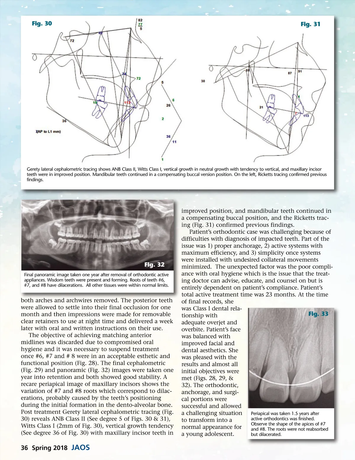
Fig. 30 Fig. 31 Gerety lateral cephalometric tracing shows ANB Class II, Witts Class I, vertical growth in neutral growth with tendency to vertical, and maxillary incisor teeth were in improved position. Mandibular teeth continued in a compensating buccal version position. On the left, Ricketts tracing confirmed previous findings. Fig. 32 Final panoramic image taken one year after removal of orthodontic active appliances. Wisdom teeth were present and forming. Roots of teeth #6, #7, and #8 have dilacerations. All other tissues were within normal limits. both arches and archwires removed. The posterior teeth were allowed to settle into their final occlusion for one month and then impressions were made for removable clear retainers to use at night time and delivered a week later with oral and written instructions on their use. The objective of achieving matching anterior midlines was discarded due to compromised oral hygiene and it was necessary to suspend treatment once #6, #7 and # 8 were in an acceptable esthetic and functional position (Fig. 28). The final cephalometric (Fig. 29) and panoramic (Fig. 32) images were taken one year into retention and both showed good stability. A recare periapical image of maxillary incisors shows the variation of #7 and #8 roots which correspond to dilac-erations, probably caused by the teeth’s positioning during the initial formation in the dento-alveolar bone. Post treatment Gerety lateral cephalometric tracing (Fig. 30) reveals ANB Class II (See degree 5 of Figs. 30 & 31), Witts Class I (2mm of Fig. 30), vertical growth tendency (See degree 36 of Fig. 30) with maxillary incisor teeth in improved position, and mandibular teeth continued in a compensating buccal position, and the Ricketts trac-ing (Fig. 31) confirmed previous findings. Patient’s orthodontic case was challenging because of difficulties with diagnosis of impacted teeth. Part of the issue was 1) proper anchorage, 2) active systems with maximum efficiency, and 3) simplicity once systems were installed with undesired collateral movements minimized. The unexpected factor was the poor compli-ance with oral hygiene which is the issue that the treat-ing doctor can advise, educate, and counsel on but is entirely dependent on patient’s compliance. Patient’s total active treatment time was 23 months. At the time of final records, she was Class I dental rela-Fig. 33 tionship with adequate overjet and overbite. Patient’s face was balanced with improved facial and dental aesthetics. She was pleased with the results and almost all initial objectives were met (Figs. 28, 29, & 32). The orthodontic, anchorage, and surgi-cal portions were successful and allowed a challenging situation Periapical was taken 1.5 years after active orthodontics was finished. to transform into a Observe the shape of the apices of #7 normal appearance for and #8. The roots were not reabsorbed a young adolescent. but dilacerated. 36 Spring 2018 JAOS
Journal of the American Orthodontic Society Spring 2018: Page 36