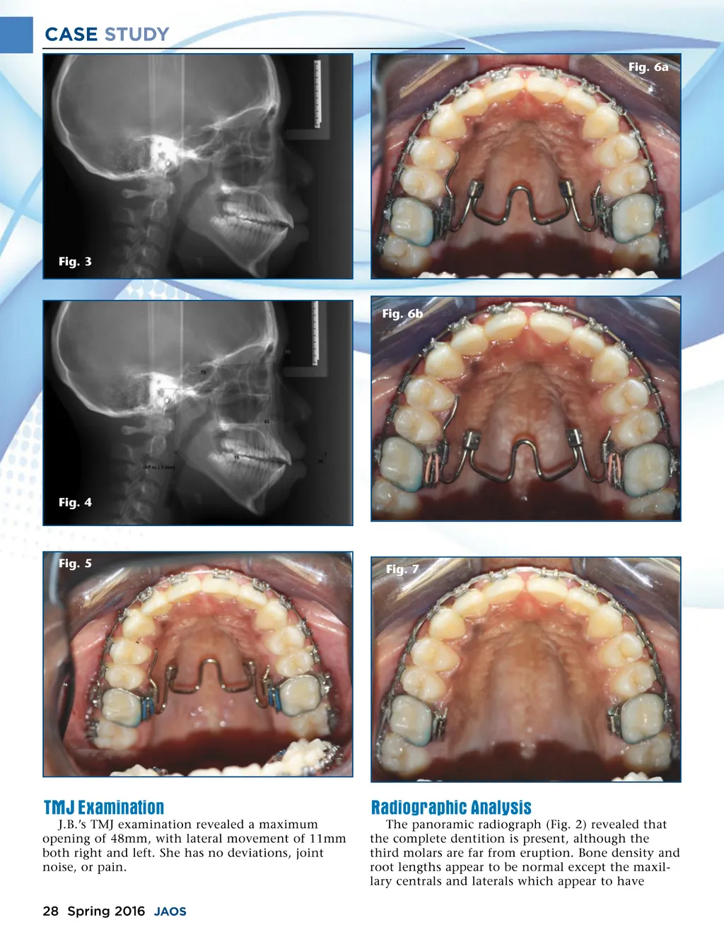
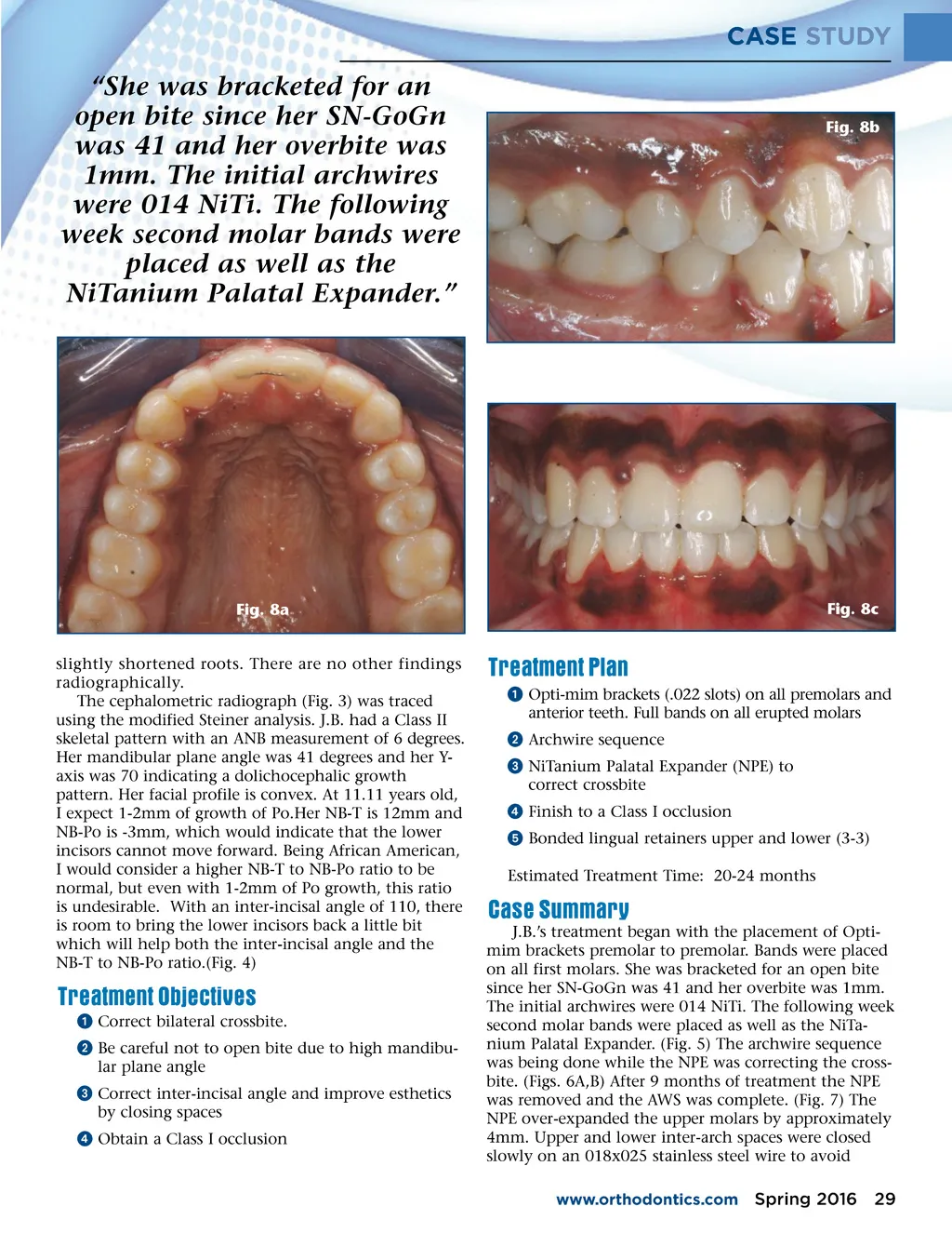
CASE STUDY Fig. 6a Fig. 3 Fig. 6b Fig. 4 Fig. 5 Fig. 7 TMJ Examination J.B.’s TMJ examination revealed a maximum opening of 48mm, with lateral movement of 11mm both right and left. She has no deviations, joint noise, or pain. Radiographic Analysis The panoramic radiograph (Fig. 2) revealed that the complete dentition is present, although the third molars are far from eruption. Bone density and root lengths appear to be normal except the maxil-lary centrals and laterals which appear to have 28 Spring 2016 JAOS
Journal of the American Orthodontic Society Spring 2016: Page 28