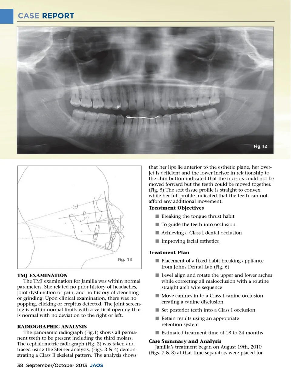
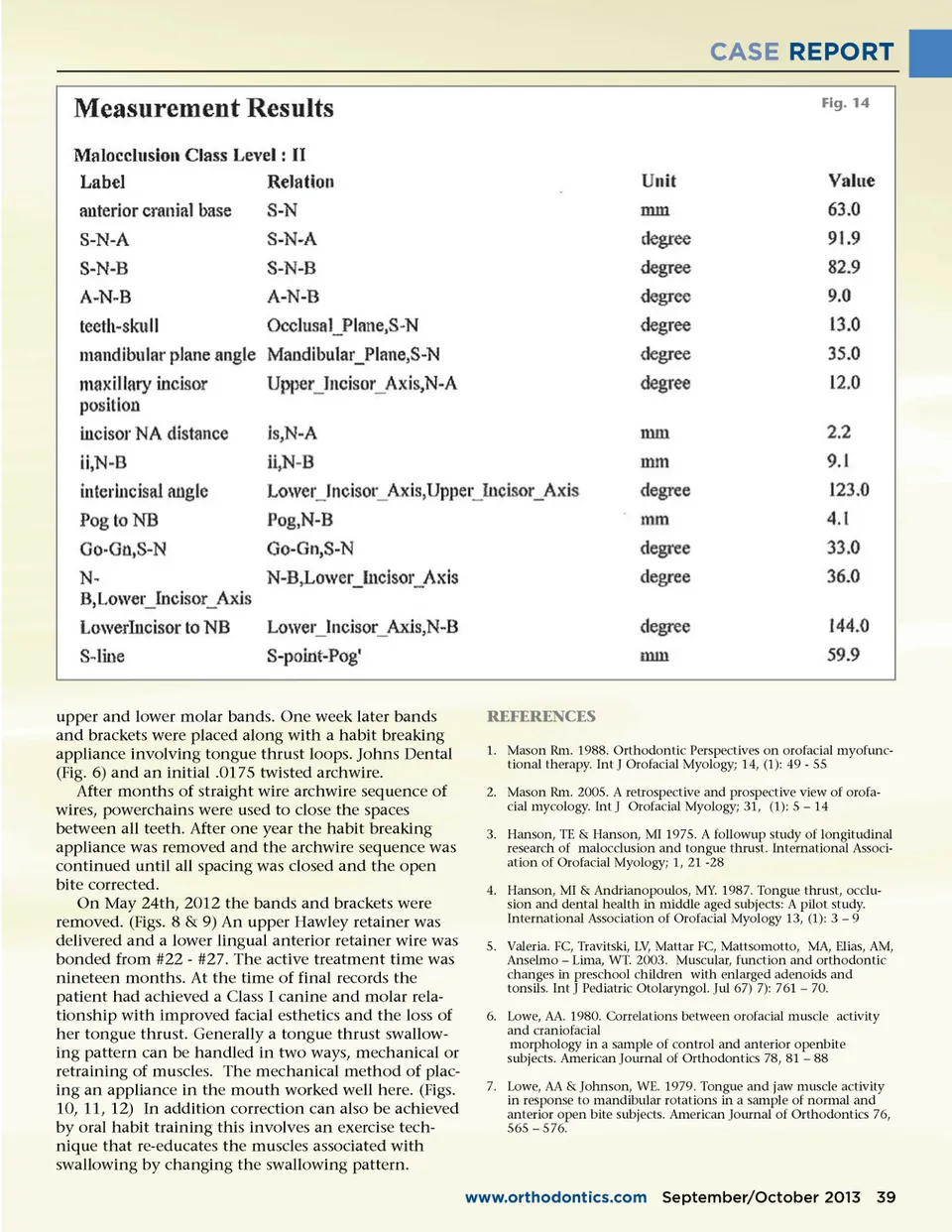
CASE REPORT Fig.12 that her lips lie anterior to the esthetic plane, her over-jet is deficient and the lower incisor in relationship to the chin button indicated that the incisors could not be moved forward but the teeth could be moved together. (Fig. 5) The soft tissue profile is straight to convex while her full profile indicated that the teeth can not afford any additional movement. Treatment Objectives í Breaking the tongue thrust habit í To guide the teeth into occlusion í Achieving a Class I dental occlusion í Improving facial esthetics Treatment Plan Fig. 13 í Placement of a fixed habit breaking appliance from Johns Dental Lab (Fig. 6) í Level align and rotate the upper and lower arches while correcting all malocclusion with a routine straight arch wire sequence í Move canines in to a Class I canine occlusion creating a canine disclusion í Set posterior teeth into a Class I occlusion í Retain results using an appropriate retention system í Estimated treatment time of 18 to 24 months Case Summary and Analysis Jamilla’s treatment began on August 19th, 2010 (Figs. 7 & 8) at that time separators were placed for TMJ EXAMINATION The TMJ examination for Jamilla was within normal parameters. She related no prior history of headaches, joint dysfunction or pain, and no history of clenching or grinding. Upon clinical examination, there was no popping, clicking or crepitus detected. The joint screen-ing is within normal limits with a vertical opening that is normal with no deviation to the right or left. RADIOGRAPHIC ANALYSIS The panoramic radiograph (Fig.1) shows all perma-nent teeth to be present including the third molars. The cephalometric radiograph (Fig. 2) was taken and traced using the Steiner analysis, (Figs. 3 & 4) demon-strating a Class II skeletal pattern. The analysis shows 38 September/October 2013 JAOS
Journal of the American Orthodontic Society September-October 2013: Page 38