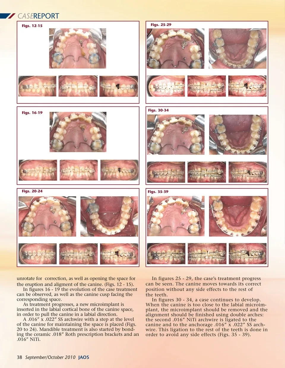
CASEREPORT Figs. 12-15 Figs. 25-29 Figs. 16-19 Figs. 30-34 Figs. 20-24 Figs. 35-39 unrotate for correction, as well as opening the space for the eruption and aligment of the canine. (Figs. 12 -15). In figures 16 -19 the evolution of the case treatment can be observed, as well as the canine cusp facing the corresponding space. As treatment progresses, a new microimplant is inserted in the labial cortical bone of the canine space, in order to pull the canine in a labial direction. A .016” x .022” SS archwire with a step at the level of the canine for maintaining the space is placed (Figs. 20 to 24). Mandible treatment is also started by bond-ing the ceramic .018” Roth prescription brackets and an .016” NiTi. 38 September/October 2010 JAOS In figures 25 -29, the case’s treatment progress can be seen. The canine moves towards its correct position without any side effects to the rest of the teeth. In figures 30 -34, a case continues to develop. When the canine is too close to the labial microim-plant, the microimplant should be removed and the alignment should be finished using double arches: the second .016” NiTi archwire is ligated to the canine and to the anchorage .016” x .022” SS arch-wire. This ligation to the rest of the teeth is done in order to avoid any side effects (Figs. 35 -39).
Journal of the American Orthodontic Society September-October 2010: Page 38