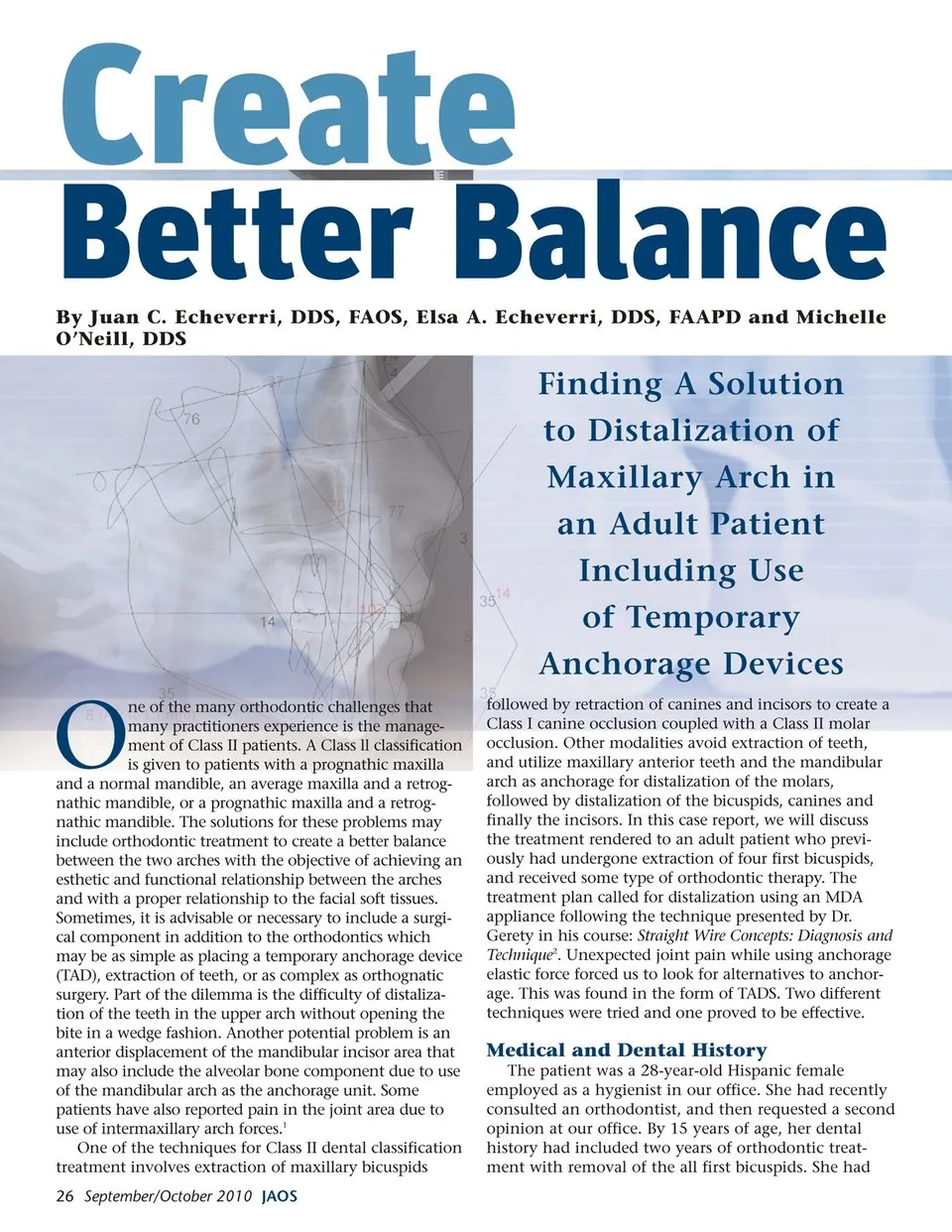
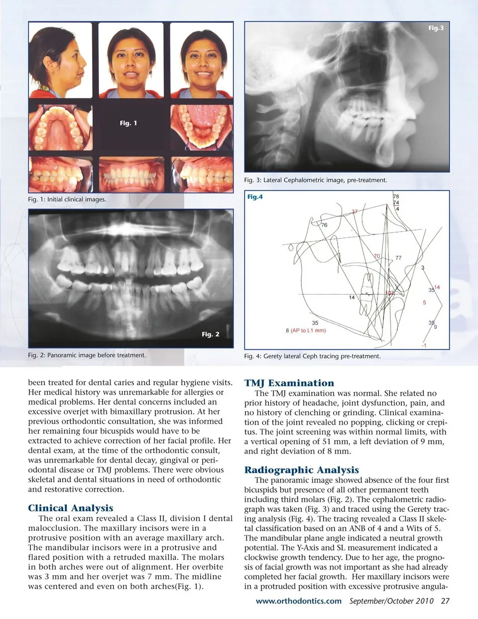
Fig.3 Fig. 1 Fig. 3: Lateral Cephalometric image, pre-treatment. Fig. 1: Initial clinical images. Fig.4 Fig. 2 Fig. 2: Panoramic image before treatment. been treated for dental caries and regular hygiene visits. Her medical history was unremarkable for allergies or medical problems. Her dental concerns included an excessive overjet with bimaxillary protrusion. At her previous orthodontic consultation, she was informed her remaining four bicuspids would have to be extracted to achieve correction of her facial profile. Her dental exam, at the time of the orthodontic consult, was unremarkable for dental decay, gingival or peri-odontal disease or TMJ problems. There were obvious skeletal and dental situations in need of orthodontic and restorative correction. Clinical Analysis The oral exam revealed a Class II, division I dental malocclusion. The maxillary incisors were in a protrusive position with an average maxillary arch. The mandibular incisors were in a protrusive and flared position with a retruded maxilla. The molars in both arches were out of alignment. Her overbite was 3 mm and her overjet was 7 mm. The midline was centered and even on both arches(Fig. 1). Fig. 4: Gerety lateral Ceph tracing pre-treatment. TMJ Examination The TMJ examination was normal. She related no prior history of headache, joint dysfunction, pain, and no history of clenching or grinding. Clinical examina-tion of the joint revealed no popping, clicking or crepi-tus. The joint screening was within normal limits, with a vertical opening of 51 mm, a left deviation of 9 mm, and right deviation of 8 mm. Radiographic Analysis The panoramic image showed absence of the four first bicuspids but presence of all other permanent teeth including third molars (Fig. 2). The cephalometric radio-graph was taken (Fig. 3) and traced using the Gerety trac-ing analysis (Fig. 4). The tracing revealed a Class II skele-tal classification based on an ANB of 4 and a Wits of 5. The mandibular plane angle indicated a neutral growth potential. The Y-Axis and SL measurement indicated a clockwise growth tendency. Due to her age, the progno-sis of facial growth was not important as she had already completed her facial growth. Her maxillary incisors were in a protruded position with excessive protrusive angula-www.orthodontics.com September/October 2010 27
Journal of the American Orthodontic Society September-October 2010: Page 27