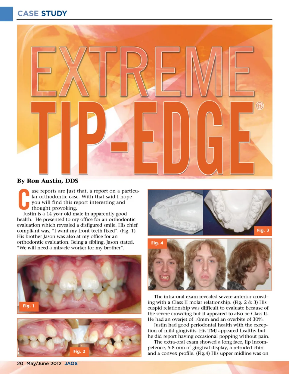
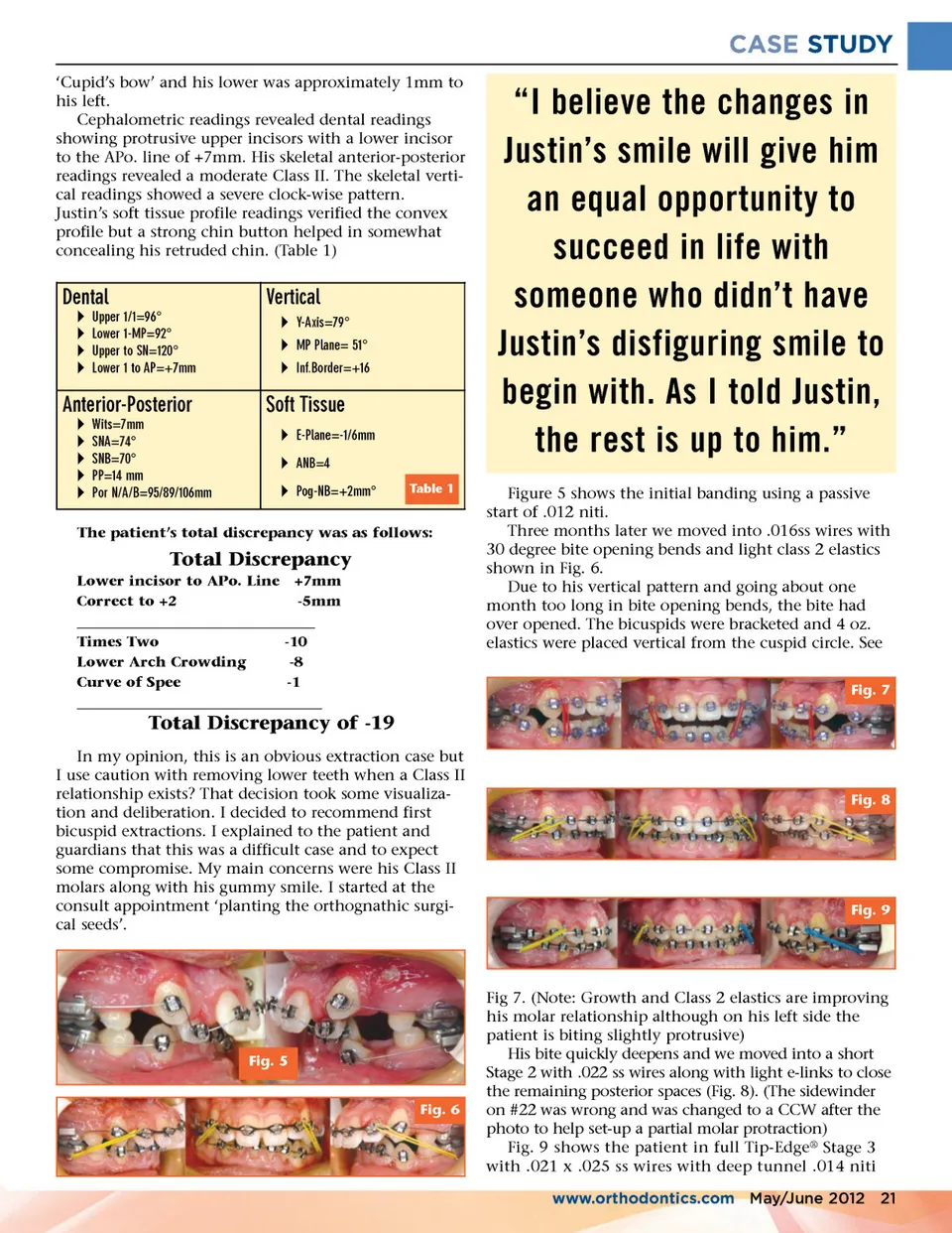
CASE STUDY ‘Cupid’s bow’ and his lower was approximately 1mm to his left. Cephalometric readings revealed dental readings showing protrusive upper incisors with a lower incisor to the APo. line of +7mm. His skeletal anterior-posterior readings revealed a moderate Class II. The skeletal verti-cal readings showed a severe clock-wise pattern. Justin’s soft tissue profile readings verified the convex profile but a strong chin button helped in somewhat concealing his retruded chin. (Table 1) Dental Ī Ī Ī Ī Upper 1/1=96° Lower 1-MP=92° Upper to SN=120° Lower 1 to AP=+7mm Vertical Ī Y-Axis=79° Ī MP Plane= 51° Ī Inf.Border=+16 Anterior-Posterior Ī Ī Ī Ī Ī Wits=7mm SNA=74° SNB=70° PP=14 mm Por N/A/B=95/89/106mm Soft Tissue Ī E-Plane=-1/6mm Ī ANB=4 Ī Pog-NB=+2mm° Table 1 “I believe the changes in Justin’s smile will give him an equal opportunity to succeed in life with someone who didn’t have Justin’s disfiguring smile to begin with. As I told Justin, the rest is up to him.” Figure 5 shows the initial banding using a passive start of .012 niti. Three months later we moved into .016ss wires with 30 degree bite opening bends and light class 2 elastics shown in Fig. 6. Due to his vertical pattern and going about one month too long in bite opening bends, the bite had over opened. The bicuspids were bracketed and 4 oz. elastics were placed vertical from the cuspid circle. See Fig. 7 The patient’s total discrepancy was as follows: Total Discrepancy Lower incisor to APo. Line +7mm Correct to +2 -5mm __________________________________ Times Two -10 Lower Arch Crowding -8 Curve of Spee -1 ___________________________________ Total Discrepancy of -19 In my opinion, this is an obvious extraction case but I use caution with removing lower teeth when a Class II relationship exists? That decision took some visualiza-tion and deliberation. I decided to recommend first bicuspid extractions. I explained to the patient and guardians that this was a difficult case and to expect some compromise. My main concerns were his Class II molars along with his gummy smile. I started at the consult appointment ‘planting the orthognathic surgi-cal seeds’. Fig. 8 Fig. 9 Fig. 5 Fig. 6 Fig 7. (Note: Growth and Class 2 elastics are improving his molar relationship although on his left side the patient is biting slightly protrusive) His bite quickly deepens and we moved into a short Stage 2 with .022 ss wires along with light e-links to close the remaining posterior spaces (Fig. 8). (The sidewinder on #22 was wrong and was changed to a CCW after the photo to help set-up a partial molar protraction) Fig. 9 shows the patient in full Tip-Edge ® Stage 3 with .021 x .025 ss wires with deep tunnel .014 niti www.orthodontics.com May/June 2012 21
Journal of the American Orthodontic Society May-June 2012: Page 21