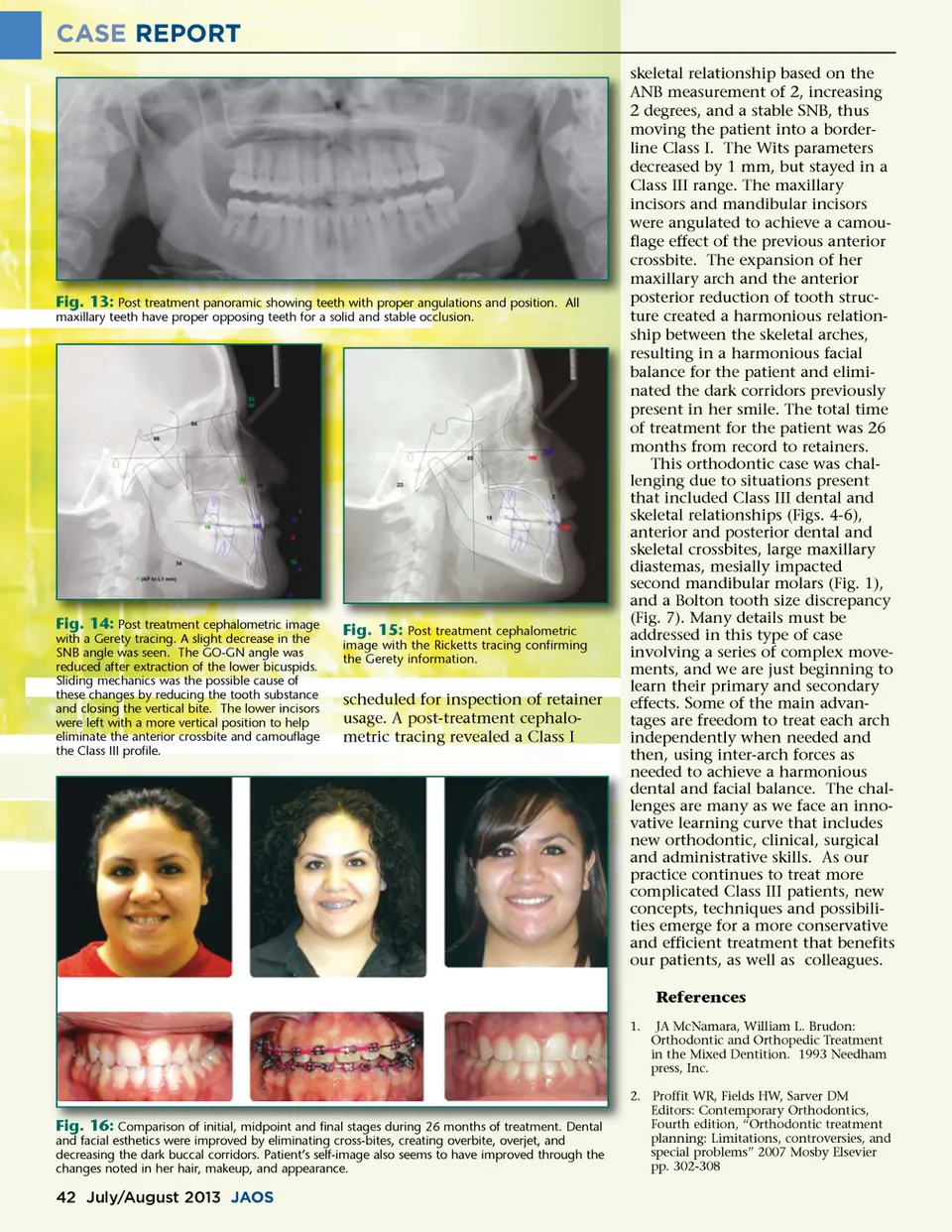
CASE REPORT skeletal relationship based on the ANB measurement of 2, increasing 2 degrees, and a stable SNB, thus moving the patient into a border-line Class I. The Wits parameters decreased by 1 mm, but stayed in a Class III range. The maxillary incisors and mandibular incisors were angulated to achieve a camou-flage effect of the previous anterior crossbite. The expansion of her maxillary arch and the anterior posterior reduction of tooth struc-ture created a harmonious relation-ship between the skeletal arches, resulting in a harmonious facial balance for the patient and elimi-nated the dark corridors previously present in her smile. The total time of treatment for the patient was 26 months from record to retainers. This orthodontic case was chal-lenging due to situations present that included Class III dental and skeletal relationships (Figs. 4-6), anterior and posterior dental and skeletal crossbites, large maxillary diastemas, mesially impacted second mandibular molars (Fig. 1), and a Bolton tooth size discrepancy (Fig. 7). Many details must be addressed in this type of case involving a series of complex move-ments, and we are just beginning to learn their primary and secondary effects. Some of the main advan-tages are freedom to treat each arch independently when needed and then, using inter-arch forces as needed to achieve a harmonious dental and facial balance. The chal-lenges are many as we face an inno-vative learning curve that includes new orthodontic, clinical, surgical and administrative skills. As our practice continues to treat more complicated Class III patients, new concepts, techniques and possibili-ties emerge for a more conservative and efficient treatment that benefits our patients, as well as colleagues. References 1. JA McNamara, William L. Brudon: Orthodontic and Orthopedic Treatment in the Mixed Dentition. 1993 Needham press, Inc. Fig. 13: Post treatment panoramic showing teeth with proper angulations and position. All maxillary teeth have proper opposing teeth for a solid and stable occlusion. Fig. 14: Post treatment cephalometric image with a Gerety tracing. A slight decrease in the SNB angle was seen. The GO-GN angle was reduced after extraction of the lower bicuspids. Sliding mechanics was the possible cause of these changes by reducing the tooth substance and closing the vertical bite. The lower incisors were left with a more vertical position to help eliminate the anterior crossbite and camouflage the Class III profile. Fig. 15: Post treatment cephalometric image with the Ricketts tracing confirming the Gerety information. scheduled for inspection of retainer usage. A post-treatment cephalo-metric tracing revealed a Class I Fig. 16: Comparison of initial, midpoint and final stages during 26 months of treatment. Dental and facial esthetics were improved by eliminating cross-bites, creating overbite, overjet, and decreasing the dark buccal corridors. Patient’s self-image also seems to have improved through the changes noted in her hair, makeup, and appearance. 2. Proffit WR, Fields HW, Sarver DM Editors: Contemporary Orthodontics, Fourth edition, “Orthodontic treatment planning: Limitations, controversies, and special problems” 2007 Mosby Elsevier pp. 302-308 42 July/August 2013 JAOS
Journal of the American Orthodontic Society July-August 2013 Buyer's Guide: Page 42