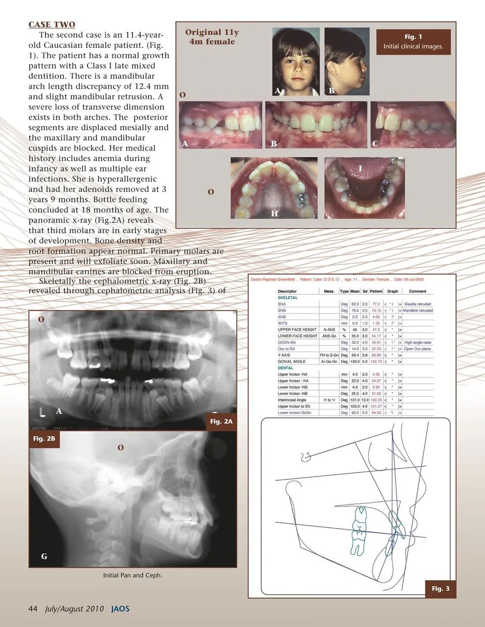
CASE TWO The second case is an 11.4-year-old Caucasian female patient. (Fig. 1). The patient has a normal growth pattern with a Class I late mixed dentition. There is a mandibular arch length discrepancy of 12.4 mm and slight mandibular retrusion. A severe loss of transverse dimension exists in both arches. The posterior segments are displaced mesially and the maxillary and mandibular cuspids are blocked. Her medical history includes anemia during infancy as well as multiple ear infections. She is hyperallergenic and had her adenoids removed at 3 years 9 months. Bottle feeding concluded at 18 months of age. The panoramic x-ray (Fig.2A) reveals that third molars are in early stages of development. Bone density and root formation appear normal. Primary molars are present and will exfoliate soon. Maxillary and mandibular canines are blocked from eruption. Skeletally the cephalometric x-ray (Fig. 2B) revealed through cephalometric analysis (Fig. 3) of O O H O Original 11y 4m female Fig. 1 Initial clinical images. AB AB I C A Fig. 2A Fig. 2B O G Initial Pan and Ceph. Fig. 3 44 July/August 2010 JAOS
Journal of the American Orthodontic Society July-August 2010/Buyer's Guide: Page 44