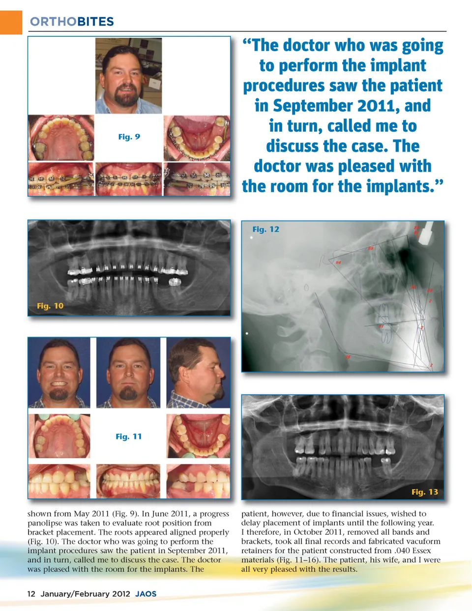
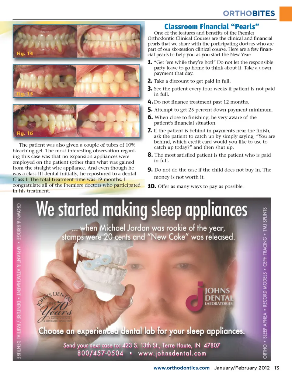
ORTHOBITES Fig. 9 “The doctor who was going to perform the implant procedures saw the patient in September 2011, and in turn, called me to discuss the case. The doctor was pleased with the room for the implants.” Fig. 12 Fig. 10 Fig. 11 Fig. 13 shown from May 2011 (Fig. 9). In June 2011, a progress panolipse was taken to evaluate root position from bracket placement. The roots appeared aligned properly (Fig. 10). The doctor who was going to perform the implant procedures saw the patient in September 2011, and in turn, called me to discuss the case. The doctor was pleased with the room for the implants. The 12 January/February 2012 JAOS patient, however, due to financial issues, wished to delay placement of implants until the following year. I therefore, in October 2011, removed all bands and brackets, took all final records and fabricated vacuform retainers for the patient constructed from .040 Essex materials (Fig. 11–16). The patient, his wife, and I were all very pleased with the results.
Journal of the American Orthodontic Society January-February 2012: Page 12