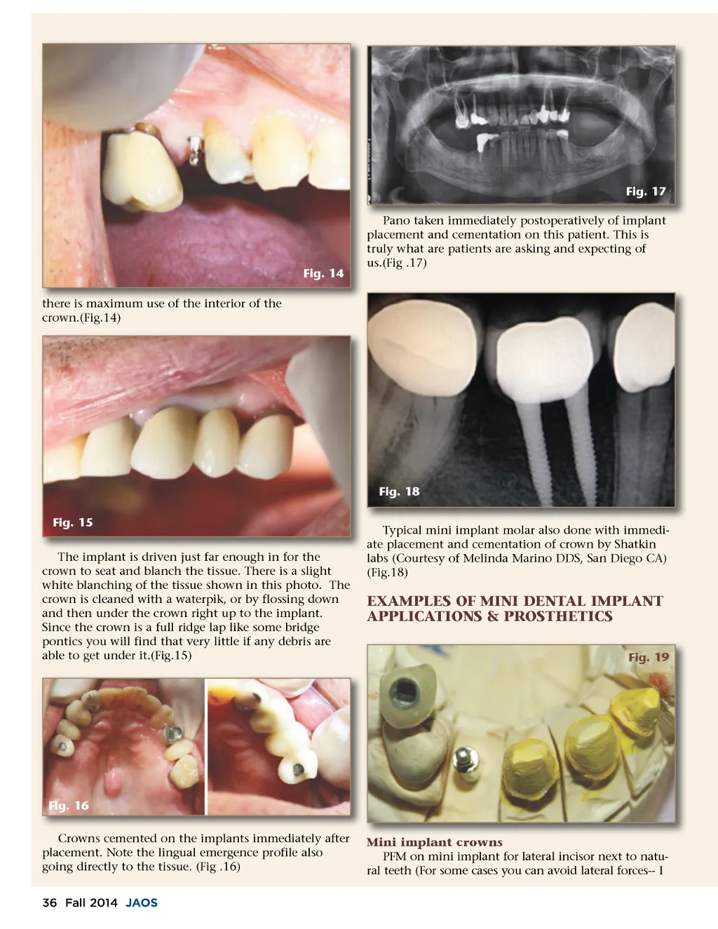
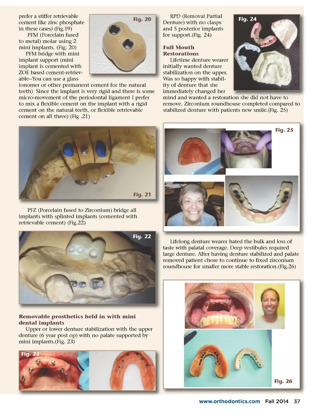
Fig. 17 Pano taken immediately postoperatively of implant placement and cementation on this patient. This is truly what are patients are asking and expecting of us.(Fig .17) Fig. 14 there is maximum use of the interior of the crown.(Fig.14) Fig. 18 Fig. 15 The is d driven just f far enough for the h implant l h in f h crown to seat and blanch the tissue. There is a slight white blanching of the tissue shown in this photo. The crown is cleaned with a waterpik, or by flossing down and then under the crown right up to the implant. Since the crown is a full ridge lap like some bridge pontics you will find that very little if any debris are able to get under it.(Fig.15) Typical mini implant molar also done with immedi-ate placement and cementation of crown by Shatkin labs (Courtesy of Melinda Marino DDS, San Diego CA) (Fig.18) EXAMPLES OF MINI DENTAL IMPLANT APPLICATIONS & PROSTHETICS Fig. 19 Fig. 16 Crowns cemented on the implants immediately after placement. Note the lingual emergence profile also going directly to the tissue. (Fig .16) 36 Fall 2014 JAOS Mi i i Mini implant l crowns PFM on mini implant for lateral incisor next to natu-ral teeth (For some cases you can avoid lateral forces--I
Journal of the American Orthodontic Society Fall 2014: Page 36